Spine Care
[Scoliosis Basics & Fundamentals 04] The Cobb Angle: How Scoliosis Severity Is Measured
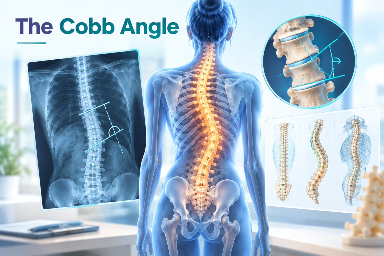
Learn how orthopedic doctors measure scoliosis severity using the Cobb angle. Discover classification tiers, measurement steps, and clinical treatment triggers. Medical diagram illustrating Cobb angle
Introduction
When diagnosing scoliosis, orthopedic surgeons and spinal specialists rely on a standardized quantitative metric to determine the degree of curvature: the Cobb angle [1]. Introduced in 1948 by American orthopedic surgeon Dr. John Robert Cobb, this measurement remains the global gold standard for assessing spinal deformities on radiographic images [1, 2].
Accurately evaluating the Cobb angle is critical not only for confirming a diagnosis of scoliosis, but also for monitoring curve progression over time and guiding clinical decision-making regarding observation, bracing, or surgical intervention [1, 3].
---
How the Cobb Angle Is Measured: Step-by-Step
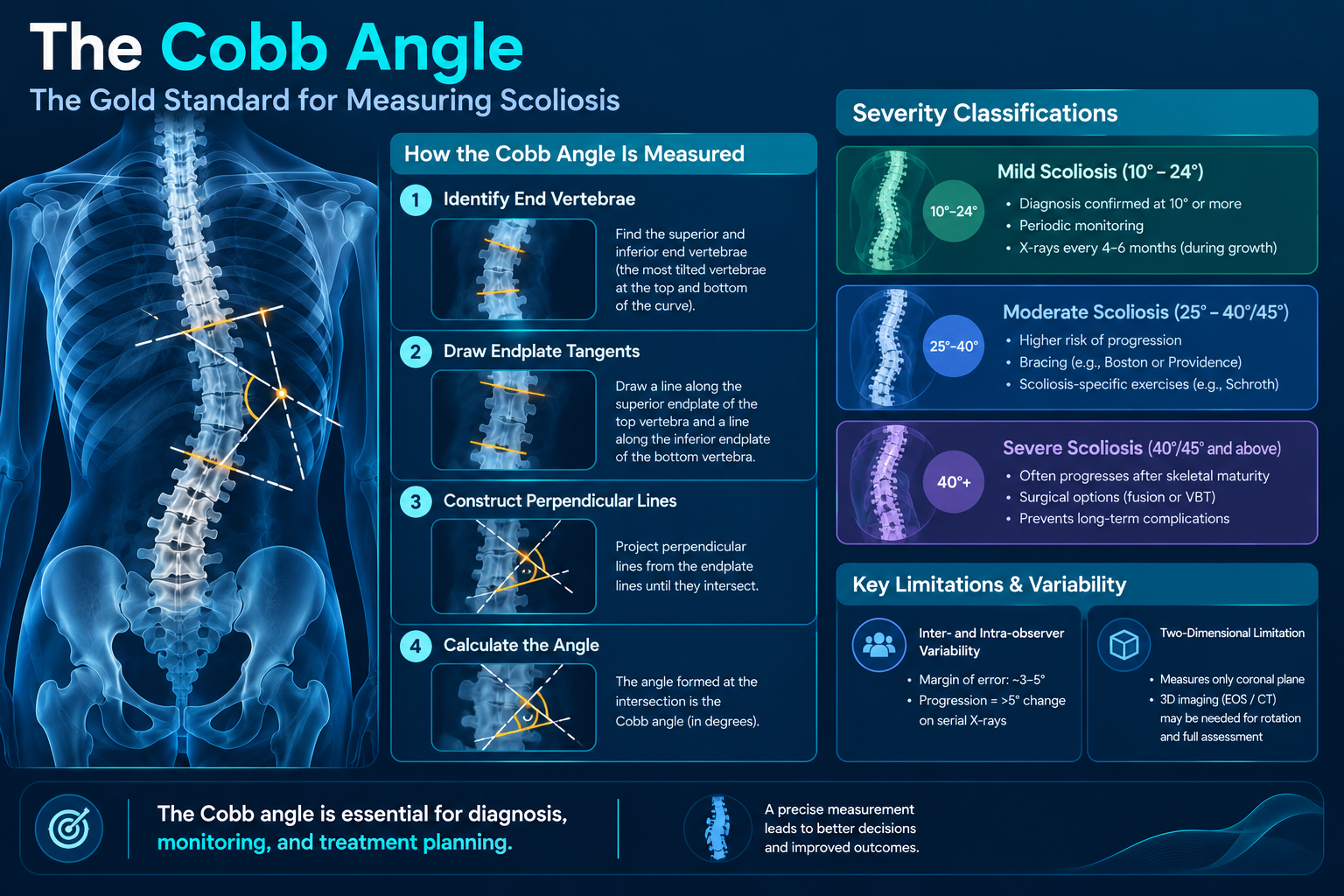
Measuring the Cobb angle requires a full-spine, standing posteroanterior (PA) X-ray [2, 3]. The measurement protocol follows four key anatomical steps [1, 2]:
- Identifying the End Vertebrae: The clinician identifies the superior and inferior end vertebrae of the scoliotic curve—the vertebrae at the top and bottom of the curve that tilt most severely toward the concavity [1, 2].
- Drawing Endplate Tangents: A straight line is drawn along the superior endplate of the top end vertebra, and another line is drawn along the inferior endplate of the bottom end vertebra [1, 2].
- Constructing Perpendicular Lines: Two perpendicular lines are projected from the endplate lines until they intersect [1].
- Calculating the Angle: The angle formed at the intersection of these two perpendicular lines represents the Cobb angle in degrees [1, 2].
---
Severity Classifications and Clinical Thresholds
In clinical practice, spinal curvature is categorized into distinct severity tiers based on the calculated Cobb angle [1, 4]:
- Mild Scoliosis (10° to 24°): Curvatures of 10 degrees or greater confirm a formal diagnosis of scoliosis [1]. Curves within this range are generally monitored through periodic clinical examinations and standing X-rays every 4 to 6 months during growth periods [1, 4].
- Moderate Scoliosis (25° to 40° or 45°): Moderate curves carry a higher risk of progression, particularly in skeletally immature adolescents [3, 4]. Non-surgical interventions, such as custom rigid bracing (e.g., Boston or Providence brace) and scoliosis-specific exercises (e.g., Schroth method), are routinely prescribed [3, 4].
- Severe Scoliosis (40° or 45° and above): Severe curvatures frequently progress even after skeletal maturity [1, 5]. Surgical options, such as posterior spinal fusion or vertebral body tethering (VBT), are typically evaluated to prevent long-term pulmonary impairment and chronic deformity [1, 5].
---
Limitations and Measurement Variability
While the Cobb angle is universally accepted, clinicians account for inherent technical variations when evaluating X-rays [2, 6]:
- Inter- and Intra-observer Variability: Studies show that manual Cobb angle measurements carry a margin of error of approximately 3 to 5 degrees between different examiners or repeated tests [2, 6]. Consequently, a curve is usually not considered to have truly progressed unless the Cobb angle increases by more than 5 degrees on serial radiographs [2, 6].
- Two-Dimensional Limitation: Scoliosis is a complex three-dimensional deformity involving lateral deviation, lordotic/kyphotic changes, and vertebral axial rotation [1, 5]. The Cobb angle measures curvature strictly in the coronal plane, meaning 3D imaging (such as EOS low-dose radiation systems or CT scans) is sometimes combined to assess vertebral rotation [5, 6].
---
Medical Disclaimer
This content is intended strictly for educational and informational purposes and does not constitute medical advice, diagnosis, or treatment. Always consult a qualified physician or orthopedic specialist for individual medical evaluation and management.
---
References & Scientific Literature
- Weinstein, S. L., Dolan, L. A., Cheng, J. C., Danielsson, A., & Morcuende, J. A. (2008). Adolescent idiopathic scoliosis. The Lancet, 371(9623), 1527-1537.
- Gstoettner, M., Sekyra, K., Walochnik, N., Winter, P., Wachter, R., & Bach, C. M. (2007). Inter- and intraobserver reliability of Cobb angle measurements in scoliosis. European Spine Journal, 16(10), 1587-1592.
- Negrini, S., Donzelli, S., Aulisa, A. G., Czaprowski, D., Schreiber, S., de Mauroy, J. C., ... & Zaina, F. (2018). 2016 SOSORT guidelines: orthopaedic and rehabilitation treatment of idiopathic scoliosis during growth. Scoliosis and Spinal Disorders, 13(1), 1-48.
- Richards, B. S., Bernstein, R. M., D'Amato, C. R., & Thompson, G. H. (2005). Standardization of terminology of the Scoliosis Research Society. Spine, 30(18), 2068-2075.
- Aebi, M. (2005). The adult scoliosis. European Spine Journal, 14(10), 925-948.
- Vrtovec, T., Pernuš, F., & Likar, B. (2009). A review of methods for quantitative evaluation of spinal curvature from medical images. European Spine Journal, 18(5), 593-607.
---