Spine Care
[Scoliosis Basics & Fundamentals 11] Congenital Scoliosis: What You Need to Know from Birth
Congenital Scoliosis: Causes, Symptoms, Diagnosis, Progression, and Treatment in Children
Learn what congenital scoliosis is, how spinal abnormalities develop before birth, what parents should know about hemivertebrae and spinal alignment, and how congenital scoliosis is diagnosed and monitored.
---
Introduction
When parents first hear that their baby or child has congenital scoliosis, one of the first questions is often, “Why did this happen?” Another is, “Will the curve get worse as my child grows?” These are understandable concerns. Congenital scoliosis can be discovered shortly after birth, during infancy, or later in childhood, sometimes before the child has any obvious symptoms. [1][2]
Congenital scoliosis is different from the much more common adolescent idiopathic scoliosis. Rather than developing without an identifiable structural cause during childhood growth, congenital scoliosis results from an abnormality in the formation or segmentation of one or more vertebrae during fetal development. The abnormal vertebral anatomy can alter the way the spine grows and may produce a persistent curvature, rotation, kyphosis, or imbalance of the trunk. [1][3]
Importantly, being born with a vertebral abnormality does not automatically mean that a child will develop severe scoliosis. Some congenital vertebral abnormalities produce little or no progression, while others can lead to significant deformity as the child grows. The risk depends on the type, number, and location of the vertebral abnormalities, as well as the amount of remaining spinal growth. [3][4]
For parents, therefore, the most important concept is not simply the number of degrees in the curve at the time of diagnosis. A relatively small curve in a very young child may require careful long-term observation if the underlying vertebral anomaly has substantial growth potential. Conversely, some structural abnormalities may remain relatively stable and require observation rather than immediate intervention. [3][4]
---
What Is Congenital Scoliosis?
Congenital scoliosis is a spinal curvature caused by a vertebral malformation that develops before birth. These abnormalities occur during embryonic development, when the vertebrae are forming and separating from one another. Depending on the developmental error, one or more vertebrae may be incompletely formed, abnormally shaped, or abnormally fused. [1][3]
The vertebral column normally develops as a series of vertebral segments that become aligned to provide structural support while allowing the spine to grow. If one part of a vertebra develops differently from another, or if adjacent vertebrae fail to separate normally, the growth of one side of the spine may differ from the other. Over time, this asymmetric growth can create or increase a spinal curve. [3][5]
This is why congenital scoliosis is best understood as a growth-related structural spinal deformity, rather than simply a posture problem. The original abnormality is present from birth, but the visible scoliosis may become more pronounced later because the child's spine continues to grow. [3][4]
---
How Is Congenital Scoliosis Different From Other Types of Scoliosis?
The word “scoliosis” describes the spinal deformity, but the underlying cause can be very different from one patient to another.
In idiopathic scoliosis, which accounts for the majority of childhood and adolescent scoliosis, no specific structural cause is identified. In congenital scoliosis, by contrast, imaging can often identify a specific vertebral malformation responsible for the abnormal spinal development. [1][6]
This distinction is important because the natural history and treatment strategy can differ substantially.
Congenital scoliosis is associated with:
- Vertebral formation defects
- Vertebral segmentation defects
- Hemivertebrae
- Wedge vertebrae
- Block vertebrae
- Unsegmented vertebral bars
- Mixed vertebral abnormalities
- Abnormal sagittal alignment or kyphosis
- Associated rib abnormalities
- Occasionally, abnormalities involving the spinal cord, heart, kidneys, or other organ systems [3][7]
Because of these differences, a child with congenital scoliosis should generally be evaluated as a developing child with a congenital spinal anomaly, rather than simply as a child with a measured Cobb angle. [2][3]
---
What Causes Congenital Scoliosis?
Abnormal Vertebral Development Before Birth
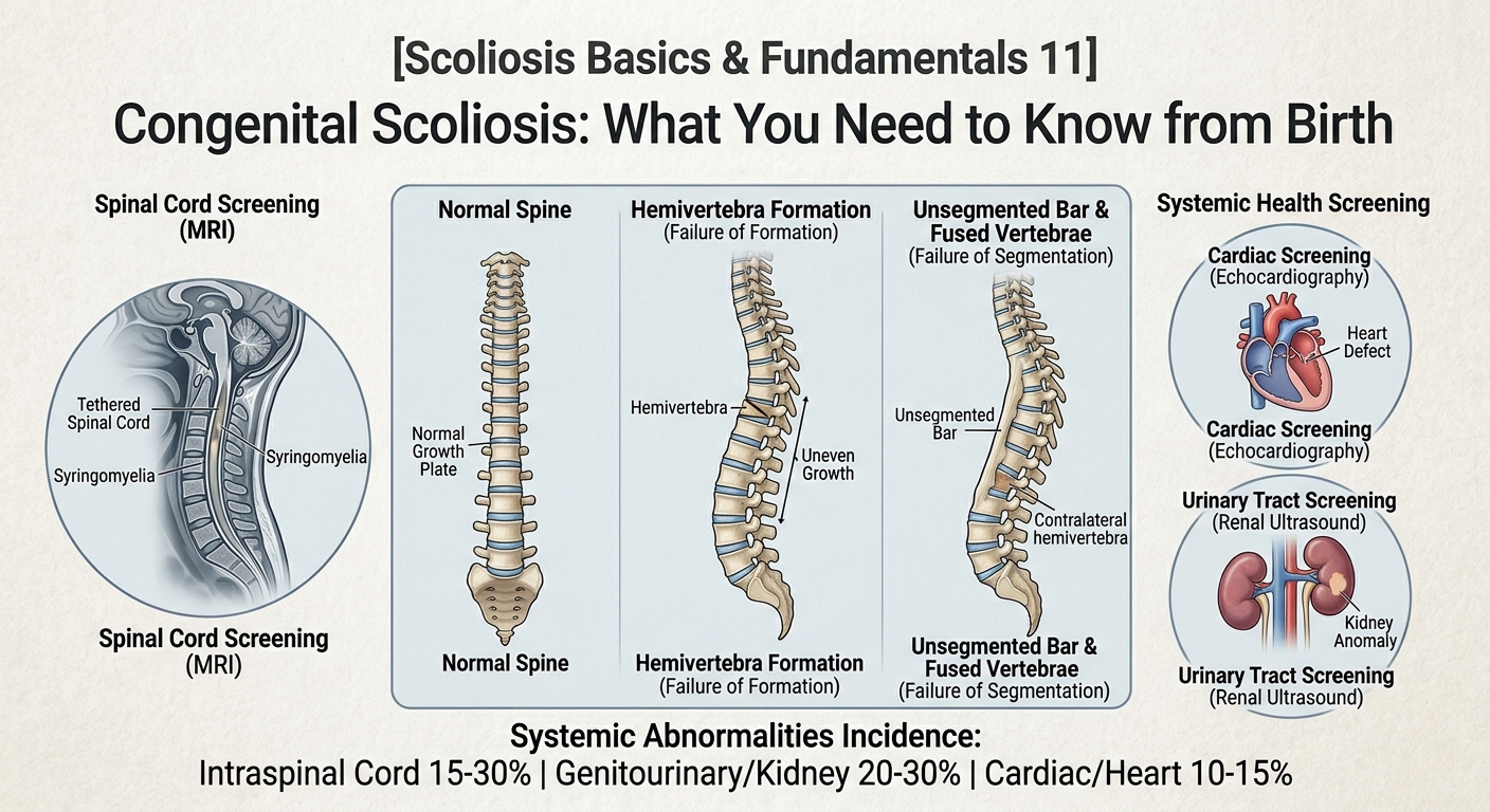
The immediate cause of congenital scoliosis is a developmental abnormality of the vertebrae during embryogenesis. The two broad categories are failure of formation and failure of segmentation, although many children have a combination of both. [3][5]
Failure of formation
A vertebra may fail to develop normally. One of the best-known examples is a hemivertebra.
A hemivertebra is a vertebra that has developed incompletely, producing a wedge-like or partially formed vertebral structure. Because one side of the vertebra may have greater or different growth potential from the other side, the spine can gradually bend toward or away from the malformed segment depending on its anatomy and location. [3][4]
Failure of segmentation
During normal development, vertebral segments separate from one another. If this separation does not occur normally, adjacent vertebrae may become partially or completely fused. A long area of fused vertebrae on one side is sometimes described as an unsegmented bar. Such a structure can restrict growth on one side while the opposite side continues to grow, potentially increasing spinal curvature. [3][4]
Mixed defects
Some children have both formation and segmentation abnormalities. One particularly important pattern is a hemivertebra on one side of the spine combined with an unsegmented bar on the opposite side. This configuration has been associated with a high risk of progression because growth is restricted asymmetrically across the spine. [3][4]
---
Is Congenital Scoliosis Hereditary?
Parents frequently worry that they caused the condition or that they passed it directly to their child.
In most cases, there is no simple explanation such as something a parent did during pregnancy causing the spinal curve. The developmental biology of congenital vertebral malformations is complex, and genetic and environmental factors have both been investigated. For many children, however, a specific cause cannot be identified. [5][8]
Congenital vertebral malformations arise during early development, and proposed mechanisms include disturbances in embryonic segmentation and vertebral formation. Research has identified possible genetic and developmental pathways, but congenital scoliosis should not generally be interpreted as evidence that a parent did something wrong during pregnancy. [5][8]
If congenital scoliosis occurs together with other congenital abnormalities or within a recognized syndrome, a pediatric specialist or geneticist may recommend additional evaluation. The need for genetic assessment depends on the individual child's clinical findings and family history. [3][8]
---
The Most Common Vertebral Abnormalities
Understanding the terminology used in an imaging report can make an overwhelming diagnosis easier for parents to understand.
Hemivertebra
A hemivertebra is a vertebra that has failed to form completely. Depending on its degree of segmentation and its location, it may have a relatively low or substantial potential to contribute to progressive scoliosis. Fully segmented hemivertebrae surrounded by relatively normal disc spaces can retain significant growth potential and may therefore be associated with progression during childhood. [3][4]
Not every hemivertebra produces severe scoliosis. The effect depends on factors such as whether it is fully or partially segmented, where it is located, whether there are additional anomalies, and whether the surrounding vertebral growth is balanced. [3][4]
Wedge Vertebra
A wedge vertebra is a vertebra that is asymmetric in shape rather than being completely absent on one side. Depending on its location and severity, it may produce a relatively mild curve or contribute to a more substantial deformity. [3][5]
Block Vertebra
A block vertebra occurs when two or more adjacent vertebrae fail to separate normally and remain fused. Some block vertebrae have relatively limited growth potential and may produce little progression of the overall curve. However, the effect depends on the location and configuration of the abnormality. [3][4]
Unsegmented Bar
An unsegmented bar is a length of fused vertebral tissue that restricts growth along one side of the spine. When the opposite side continues to grow, the imbalance can contribute to progressive curvature. Unilateral unsegmented bars, especially when combined with a contralateral hemivertebra, are considered important high-risk configurations for progression. [3][4]
---
Why Can Congenital Scoliosis Get Worse as a Child Grows?
This is one of the most important questions for parents.
The answer is related to asymmetric spinal growth.
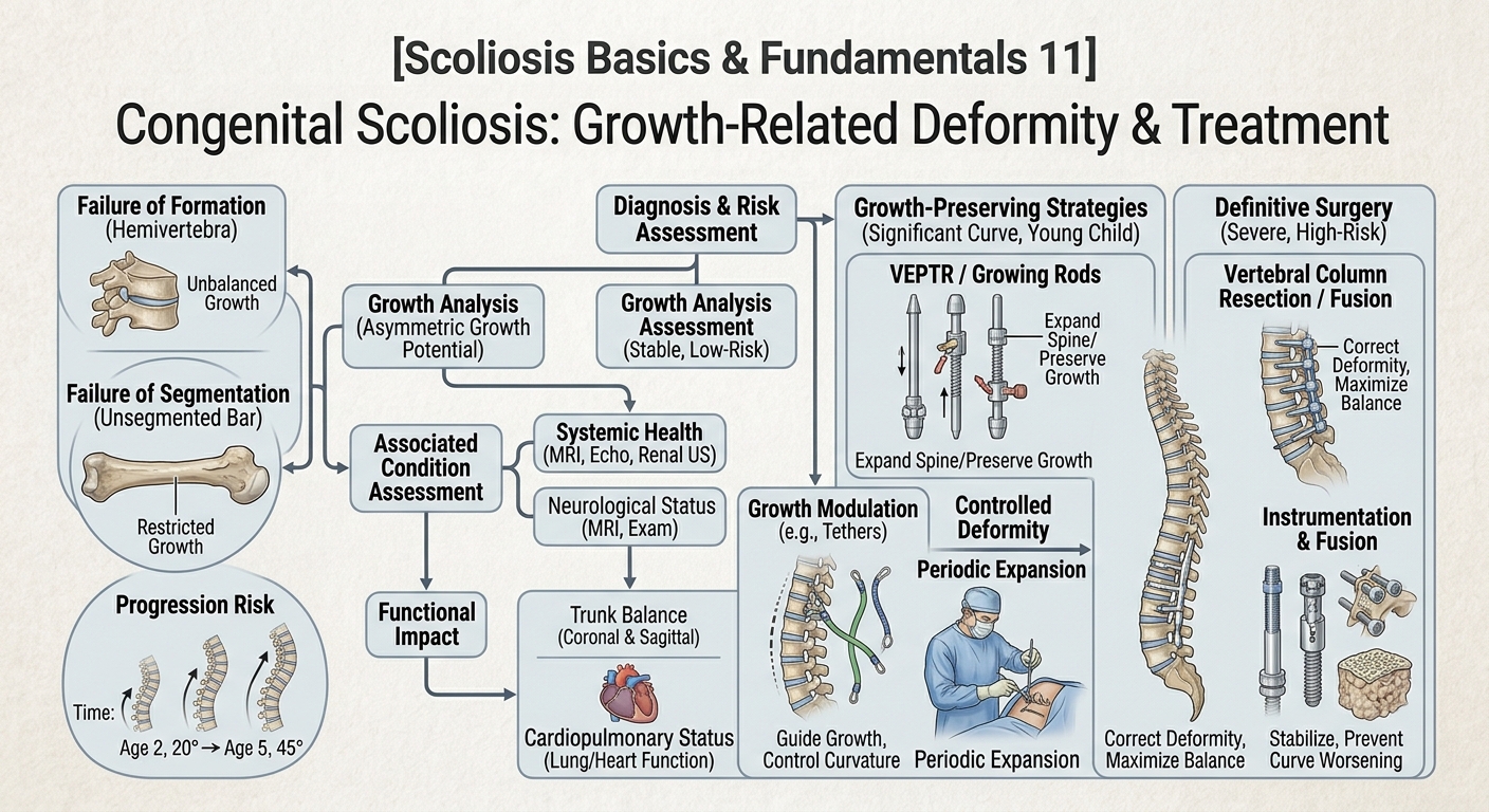
Imagine a young child's spine as a growing structure. If one side of the spine has normal growth potential while the opposite side is restricted by a malformed vertebra or bony bar, the two sides may not grow at the same rate. As growth continues, the difference can gradually increase the curvature. [3][4]
This means that a child can be born with a vertebral abnormality but initially have only a modest visible curve. The deformity may become more obvious later because the underlying structural imbalance interacts with normal growth. [3][4]
The rate of progression is highly variable. Studies indicate that approximately half of congenital curves may progress relatively quickly, while others progress more slowly or remain relatively stable. The risk depends heavily on the anomaly pattern, location, age, and remaining growth potential. [3][4]
Periods of particular concern include:
- Early childhood, particularly the first several years of life
- Periods of rapid spinal growth
- The adolescent growth spurt
- Situations involving multiple vertebral anomalies
- Mixed formation and segmentation defects
- Thoracolumbar deformities
- Curves associated with significant kyphosis or trunk imbalance [3][4]
Therefore, a small Cobb angle does not necessarily mean a low-risk congenital scoliosis. The underlying vertebral anatomy is essential to understanding prognosis. [3][4]
---
Understanding the Cobb Angle in Congenital Scoliosis
The Cobb angle is commonly used to quantify the magnitude of a spinal curve on an X-ray.
The measurement is obtained by identifying the vertebrae at the upper and lower limits of the curve and calculating the angle formed by the relevant endplate lines. A Cobb angle of approximately 10 degrees or greater is conventionally used to define scoliosis radiographically. [9][10]
For parents, however, it is important to understand that the Cobb angle is one measurement, not the entire diagnosis.
For example, two children may both have a 25-degree curve, but their long-term outlook could be very different. One child might have a relatively stable vertebral configuration with limited remaining growth potential, while another might have a fully segmented hemivertebra and an opposing unsegmented bar with substantial growth remaining. [3][4]
When evaluating congenital scoliosis, clinicians therefore consider:
- Cobb angle
- Vertebral anomaly type
- Number of abnormal vertebrae
- Location of the abnormality
- Degree of vertebral segmentation
- Kyphosis or lordosis
- Vertebral rotation
- Coronal balance
- Sagittal balance
- Trunk shift
- Pelvic alignment
- Skeletal maturity
- Remaining growth
- Neurological findings
- Associated congenital abnormalities [3][7]
---
Why Spinal Alignment Matters
Congenital scoliosis can affect more than the appearance of the spine.
A significant congenital deformity can alter coronal spinal alignment, causing the trunk to shift to one side. It can also affect sagittal alignment, particularly when congenital kyphosis or kyphoscoliosis is present. In some children, vertebral rotation further changes the three-dimensional shape of the thorax and trunk. [3][4]
Parents may notice:
- One shoulder higher than the other
- Uneven shoulder blades
- An asymmetric waist
- One side of the rib cage appearing more prominent
- A tilted or shifted trunk
- Uneven pelvic position
- A visible spinal curve
- Increasing difficulty maintaining a balanced posture
These visible changes are useful clues, but they cannot determine the severity or progression risk by themselves. Radiographic evaluation is necessary to understand the underlying vertebral anatomy. [1][3]
---
What Other Health Problems Can Be Associated With Congenital Scoliosis?
One of the most important differences between congenital and idiopathic scoliosis is that congenital vertebral abnormalities can occur alongside abnormalities in other organ systems.
These may involve the:
- Spinal cord and nervous system
- Heart and cardiovascular system
- Kidneys and urinary tract
- Ribs and chest wall
- Other congenital structures [7][11]
This does not mean that every child with congenital scoliosis has another health problem. It means that the possibility is important enough that clinicians may recommend screening even when the child appears healthy.
Studies of children with congenital scoliosis have found substantial rates of associated intraspinal, cardiac, and genitourinary abnormalities. The exact percentages vary considerably between studies and patient populations, but the evidence supports comprehensive evaluation rather than assuming the vertebral abnormality is isolated. [11][12]
---
Why Might a Child Need an MRI?
An MRI of the spine provides information that a standard X-ray cannot.
X-rays are excellent for evaluating bony alignment, vertebral shape, and measuring the Cobb angle. MRI, however, can show the spinal cord, nerve structures, discs, and other soft tissues. [7][13]
In congenital scoliosis, MRI may identify associated conditions such as:
- Tethered spinal cord
- Syringomyelia
- Diastematomyelia
- Other spinal dysraphism
- Abnormalities of the spinal canal
- Other neural-axis abnormalities [3][7]
These abnormalities may sometimes exist without obvious neurological symptoms. This is one reason why specialist evaluation may recommend MRI even when a child appears neurologically normal. [7][11]
MRI is particularly important when there are neurological findings, complex congenital anomalies, significant kyphosis, or when corrective spinal surgery is being considered. The exact imaging protocol and timing should be determined by the child's treating specialists. [3][7]
---
Why Are the Heart and Kidneys Sometimes Examined?
The same developmental processes that affect vertebral formation can sometimes be associated with abnormalities in other organ systems.
For this reason, children with congenital scoliosis may undergo investigations such as:
- Echocardiography to evaluate the heart
- Renal ultrasound to examine the kidneys and urinary tract
- Additional abdominal or systemic imaging when clinically indicated [7][11]
Research has documented significant rates of cardiac and genitourinary abnormalities in children with congenital scoliosis. One large retrospective study reported intraspinal, urogenital, and cardiac abnormalities at substantial frequencies, supporting a comprehensive diagnostic approach. [11]
This can be unsettling for parents, particularly when their child has no symptoms. However, the purpose of screening is precisely to identify abnormalities that may otherwise remain unnoticed and to ensure that any necessary treatment or precautions are addressed early. [11][12]
---
What Symptoms Might Parents Notice?
Congenital scoliosis may cause no symptoms at all during early childhood.
Some children are diagnosed incidentally when an X-ray is obtained for another reason. Others are identified because a parent, pediatrician, or school screening program notices asymmetry. [1][3]
Possible physical signs include:
- Uneven shoulders
- Uneven waistline
- Prominent shoulder blade
- Rib prominence
- Trunk leaning or shifting to one side
- Uneven hips or pelvis
- Visible curvature of the spine
- Abnormal posture symmetry
- Increasing back or neck deformity
- Shortening or imbalance of the trunk in severe cases [3][4]
Pain is not necessarily the first or most prominent symptom in young children with congenital scoliosis. Therefore, waiting for pain before seeking evaluation is not appropriate when a persistent spinal asymmetry is visible. [1][3]
---
How Is Congenital Scoliosis Diagnosed?
Diagnosis usually begins with a medical history and physical examination followed by appropriate imaging.
Step 1: Clinical Examination
A clinician may evaluate:
- Shoulder and pelvic symmetry
- Trunk balance
- Spinal contour
- Rib prominence
- Range of motion
- Neurological function
- Skin findings suggesting an underlying spinal anomaly
- Gait and overall motor function [1][3]
The Adam's forward bend test may reveal trunk or rib asymmetry, although it is a screening examination rather than a definitive diagnostic test. [1]
Step 2: Standing Spinal X-rays
Standing radiographs allow clinicians to evaluate the overall spinal alignment and calculate the Cobb angle.
The X-ray can also reveal structural abnormalities such as hemivertebrae, fused vertebrae, segmentation defects, and abnormal curvature patterns. [3][9]
Step 3: MRI
MRI may be used to evaluate the spinal cord and neural axis, particularly when congenital anomalies are complex or when neurological abnormalities or surgical planning make detailed evaluation important. [3][7]
Step 4: Additional Imaging
In complex congenital deformities, CT—sometimes with three-dimensional reconstruction—can provide detailed information about vertebral anatomy and may be particularly useful for surgical planning. Because CT uses ionizing radiation, its use in children should be justified according to the clinical situation rather than performed routinely without indication. [7]
---
Does Every Child With Congenital Scoliosis Need Surgery?
No.
The discovery of congenital scoliosis does not automatically mean that surgery is necessary.
Some curves can be observed with periodic clinical and radiographic follow-up, particularly when the vertebral abnormality has a relatively low progression potential and the spinal alignment remains balanced. [3][4]
The decision depends on factors such as:
- Current curve magnitude
- Rate of progression
- Vertebral anomaly type
- Child's age
- Remaining growth
- Curve location
- Kyphosis or other sagittal deformity
- Trunk balance
- Neurological findings
- Associated abnormalities
- Respiratory or functional consequences [3][7]
When surgery is considered, the goal is generally not simply to make an X-ray “straight.” The broader objectives include preventing progressive deformity, maintaining or improving coronal and sagittal balance, preserving spinal and thoracic growth when possible, and protecting neurological and cardiopulmonary function. [3][7]
---
What Treatments May Be Considered?
Treatment must be individualized.
Observation
Observation can be appropriate for selected children with relatively stable congenital abnormalities. Follow-up is important because the curve may change as the child grows. Serial assessments allow clinicians to identify progression before the deformity becomes severe. [3][4]
Bracing
Bracing has a more limited role in congenital scoliosis than in adolescent idiopathic scoliosis because congenital curves are often associated with rigid structural abnormalities. A brace cannot remove or correct the underlying malformed vertebra. However, in selected cases it may be considered for associated compensatory curves or specific growth-related circumstances. [3][4]
Parents should therefore be cautious about assuming that a brace used for adolescent idiopathic scoliosis will have the same role in congenital scoliosis.
Growth-Preserving Strategies
For very young children with significant progressive deformity, specialists may consider growth-friendly approaches designed to control spinal deformity while allowing continued growth of the spine and thorax. The appropriate strategy depends heavily on the child's age, anatomy, curve pattern, and surgical needs. [7]
Surgery
Surgery may be considered when there is significant or progressive deformity, particularly when the underlying anomaly has a high progression potential.
Procedures can include techniques designed to address the malformed vertebra, stabilize the spine, correct the deformity, or control progression while considering remaining growth. The specific operation varies considerably from child to child. [3][7]
Because congenital spinal anatomy can be complex, treatment is generally best planned by a pediatric spine or pediatric spinal deformity specialist with experience in congenital conditions. [3][7]
---
What Should Parents Ask the Spine Specialist?
A diagnosis can feel overwhelming. Parents may find it helpful to bring a written list of questions to the appointment.
Consider asking:
About the anatomy
- What type of vertebral abnormality does my child have?
- Is there a hemivertebra?
- Is there an unsegmented bar?
- Are the vertebrae partially or completely fused?
- Where is the abnormality located?
About progression
- What is the current Cobb angle?
- Is the curve changing over time?
- How much growth does my child have remaining?
- Does the particular vertebral anomaly have a high risk of progression?
- Is there kyphosis or significant trunk imbalance?
About associated conditions
- Does my child need a spinal MRI?
- Should the heart be evaluated?
- Should the kidneys and urinary tract be screened?
- Are there any neurological findings that require further investigation?
About treatment
- Is observation appropriate?
- How often should follow-up X-rays be performed?
- Would bracing have a meaningful role?
- If surgery is recommended, what is the objective?
- How will treatment affect future spinal and thoracic growth?
Having these questions answered can help parents understand not only what the spine looks like today, but also what the medical team expects may happen during growth.
---
Important Precautions and Medical Disclaimer
The information provided by 4scoliosis.com is intended for educational purposes and should not replace professional medical assessment, diagnosis, or individualized treatment. Congenital scoliosis is a heterogeneous condition, and two children with apparently similar Cobb angles may have very different underlying vertebral anatomy and progression risks. [3][7]
A child with congenital scoliosis should not be evaluated solely on the basis of the Cobb angle. The vertebral anomaly, skeletal maturity, curve location, spinal balance, neurological status, and associated congenital abnormalities are all important considerations. [3][4]
Parents should seek prompt medical evaluation if a child's spinal deformity appears to be progressing rapidly or if the child develops neurological symptoms such as weakness, numbness, changes in walking, loss of coordination, or problems with bowel or bladder control. Breathing difficulties or significant changes in chest development also warrant medical attention. [3][7]
Parents should also avoid assuming that an exercise program, brace, manual therapy, or other non-surgical intervention can correct the underlying congenital vertebral malformation. Some interventions may have a role in selected patients, but treatment should be based on the child's specific anatomy and progression risk. [3][7]
This article does not provide an individualized diagnosis or recommend surgery, bracing, medication, exercise, manual therapy, or any other treatment for a specific child. Decisions about imaging and treatment should be made with qualified healthcare professionals familiar with pediatric spinal deformity.
---
Conclusion: What Parents Should Remember
Congenital scoliosis begins with a vertebral developmental abnormality that occurs before birth. The abnormality may involve failure of vertebral formation, failure of segmentation, or a combination of both. Hemivertebrae, wedge vertebrae, fused vertebrae, and unsegmented bars are among the structural abnormalities that can contribute to congenital spinal curvature. [3][5]
The presence of a congenital vertebral anomaly does not automatically mean that severe scoliosis will develop. Some children remain relatively stable, while others experience significant progression as the spine grows. The risk depends on the specific anatomy, location and number of anomalies, remaining growth, and the balance between the two sides of the developing spine. [3][4]
For this reason, early and accurate evaluation is important. A child's assessment may involve standing X-rays and Cobb-angle measurement, but it may also require MRI of the spine, evaluation of the heart and kidneys, and other investigations depending on the clinical situation. [7][11]
For parents, perhaps the most important message is this:
A diagnosis of congenital scoliosis is not simply a measurement on an X-ray. It is a developmental condition that must be understood in the context of the child's anatomy, growth, spinal alignment, and overall health.
If your child has been diagnosed with congenital scoliosis, ask the treating specialist to explain the exact vertebral abnormality, its estimated progression risk, the current Cobb angle, the child's remaining growth potential, and whether associated spinal cord, cardiac, or renal abnormalities have been evaluated.
Regular follow-up is important because the spine is still growing. Detecting meaningful progression early can give the medical team more options for preserving spinal balance, thoracic development, and overall function. [3][7]
At 4scoliosis.com, our goal is to help parents and patients understand scoliosis in clear, evidence-informed language. A diagnosis can be frightening, but understanding the condition is an important first step toward making informed decisions about a child's spinal health.
---
References
[1] Scoliosis Research Society. Scoliosis: Patient and Family Information. Scoliosis Research Society – Scoliosis Information
[2] Hwang SW, Samdani AF, Jea A, et al. A comprehensive review of the diagnosis and management of congenital scoliosis. Child's Nervous System. 2018;34:2151–2160. PMID: 30078055.
[3] Giampietro PF, Raggio CL, Reynolds C, et al. Congenital scoliosis: an up-to-date. Journal/PMC review of congenital spinal deformity and natural history.
[4] Tello CA, et al. Congenital scoliosis: a narrative review and proposal of a treatment algorithm. Journal of Orthopaedics and Traumatology. 2022.
[5] Giampietro PF, Raggio CL, Reynolds C, et al. Clinical, Genetic and Environmental Factors Associated with Congenital Vertebral Malformations.
[6] American Academy of Orthopaedic Surgeons. Scoliosis in Children.
[7] Current Concepts – Congenital Scoliosis. Review of diagnostic evaluation, MRI, associated visceral abnormalities, and treatment considerations.
[8] Congenital and Idiopathic Scoliosis: Clinical and Genetic Aspects. Review of vertebral formation and segmentation defects and their developmental basis.
[9] Cobb JR. Outline for the Study of Scoliosis. Instructional Course Lectures. 1948;5:261–275.
[10] Langensiepen S, Semler O, Sobottke R, et al. Measuring procedures to determine the Cobb angle in idiopathic scoliosis: a systematic review. European Spine Journal. 2013;22:2360–2371.
[11] Bas T, et al. The Incidence and Interrelationship of Concomitant Anomalies in Congenital Scoliosis. Turkish Neurosurgery. PMID: 30649818.
[12] Burnei G, et al. Congenital scoliosis and associated systemic abnormalities. Review of cardiac, genitourinary, and other associated anomalies.
[13] Congenital spinal deformity: a comprehensive assessment at presentation. Study evaluating intraspinal, cardiac, and urogenital abnormalities associated with congenital spinal deformity.
[14] Incidence of cardiac anomalies in congenital vertebral deformity: systematic review and meta-analysis of 2910 patients. Meta-analysis of cardiac abnormalities associated with congenital vertebral deformity.
[15] Hemivertebra Resection in Small Children: A Literature Review. Review of congenital scoliosis associated with hemivertebrae and treatment considerations in young children.