Spine Care
[Biomechanics, Tensegrity & Meridian Concepts 13] The Impact of Rib Hump Deformation on Thoracic Biomechanics
Rib hump deformity reflects three-dimensional spinal rotation and can alter thoracic mechanics, load distribution, chest wall motion, and respiratory function.

Introduction
A rib hump is one of the most visible physical signs of thoracic scoliosis. When a person bends forward, one side of the posterior rib cage may become more prominent, creating an asymmetrical “hump.” The Scoliosis Research Society describes this rib prominence as a consequence of the rotational component of scoliosis rather than simply a side-to-side spinal curve. [1]
Although the rib hump is often discussed as a cosmetic feature, its biomechanical significance is much broader. The ribs, vertebrae, sternum, costal cartilage, respiratory muscles, and surrounding soft tissues form an integrated three-dimensional structure. Changes in one component can influence the mechanical behavior of the others.
In thoracic scoliosis, vertebral rotation can change the orientation of the ribs. The altered ribs can then modify the shape, stiffness, and movement of the thoracic cage. This creates a complex mechanical environment in which spinal loading, rib motion, respiratory mechanics, and muscle activity may become asymmetrical. [2,3]
Understanding this relationship is important because the rib hump provides information about the three-dimensional nature of scoliosis that cannot be captured by the Cobb angle alone.
1. What Is a Rib Hump?
A rib hump is a visible posterior prominence of the ribs, most commonly observed during the forward-bending or Adams test. It is particularly associated with thoracic curves and reflects axial rotation of the vertebrae and associated deformation of the rib cage. [1]
The key point is that scoliosis is not simply a lateral deviation.
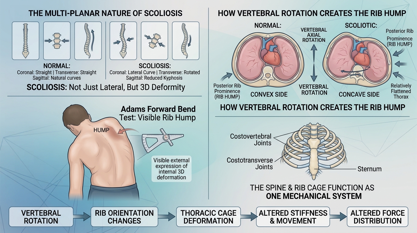
A typical structural scoliosis involves changes in multiple planes:
- Coronal plane: lateral curvature
- Sagittal plane: changes in thoracic kyphosis or lordosis
- Transverse plane: vertebral rotation
The rib hump is largely an external manifestation of the transverse-plane component.
This explains why two patients with similar Cobb angles can have noticeably different trunk and rib-cage appearances.
Clinical concept: Cobb angle describes one important aspect of spinal deformity, while rib prominence provides additional information about three-dimensional trunk rotation.
2. The Spine and Rib Cage Function as One Mechanical System
The thoracic spine cannot be considered mechanically independent from the rib cage.
The ribs attach to the thoracic vertebrae through costovertebral and costotransverse joints and connect anteriorly through the sternum and costal cartilage. Together, these structures create the rib-vertebral-sternal complex.
Biomechanical studies indicate that the intact rib cage contributes substantially to thoracic spinal stability. A systematic review found that the rib cage reduces thoracic spinal range of motion and neutral-zone motion while increasing stiffness and resistance to compression, particularly in axial rotation. [2]
This means that changes in rib geometry can potentially influence how the thoracic spine responds to external forces.
The mechanical relationship can be represented as:
Vertebral rotation → rib orientation changes → thoracic cage deformation → altered stiffness and movement → altered force distribution
This is not necessarily a simple one-way process. The spine and rib cage can influence each other as part of an interconnected mechanical system.
3. How Vertebral Rotation Produces Rib Prominence
In thoracic scoliosis, vertebral bodies rotate around the longitudinal axis of the spine.
Because the ribs are mechanically connected to the vertebrae, this rotation changes their orientation.
On the convex side, posterior rib prominence may become more pronounced, producing the visible rib hump. At the same time, the opposite side of the thorax may appear relatively flattened or compressed.
The result is not merely a curved spine but a distorted three-dimensional thoracic cage.
This distinction is important when evaluating scoliosis because a radiograph viewed only from the front cannot fully represent the rotational and rib-cage components of the deformity.
4. Rib Hump and Three-Dimensional Thoracic Deformation
The thoracic cage normally has a relatively symmetrical three-dimensional configuration that permits both stability and respiratory movement.
With progressive thoracic deformity, this geometry can change.
Research using three-dimensional imaging has demonstrated differences in the morphology of the rib cage and lungs between individuals with adolescent idiopathic scoliosis and healthy controls. [3]
Changes may include:
- asymmetric rib orientation
- altered thoracic width
- changes in anteroposterior dimensions
- altered rib curvature
- asymmetric lung spaces
- changes in the relationship between ribs and vertebrae
Consequently, the visible rib hump can be viewed as the external expression of a deeper three-dimensional deformation.
5. How Rib Hump Deformation Changes Mechanical Loading
The thoracic cage participates in transmitting and distributing forces around the spine.
When its geometry becomes asymmetric, mechanical loads may no longer be distributed evenly.
Potentially affected forces include:
- axial compression
- bending moments
- rotational moments
- shear forces
- rib–vertebral joint forces
- muscle-generated forces
Finite-element studies have investigated the effect of the rib cage on the static characteristics of scoliotic spines and found that including the rib cage changes the mechanical behavior of the spinal model under different loading conditions. [4]
This is important because a spine model that ignores the rib cage may not accurately represent the mechanical environment of a real thoracic spine.
6. Rib Hump and Axial Rotation
Axial rotation is particularly important in understanding rib hump deformity.
The Cobb angle primarily describes coronal-plane curvature. Vertebral axial rotation describes another dimension of the deformity.
The Scoliosis Research Society's terminology specifically defines vertebral axial rotation as rotation of a vertebra around its local longitudinal axis. [5]
The rib cage translates this internal vertebral rotation into a visible external change.
Therefore:
Cobb angle ≠ complete measurement of rib-cage deformity
A patient can have a moderate Cobb angle but relatively substantial trunk rotation, while another patient with a similar Cobb angle may have less visible rib prominence.
This is one reason clinical assessment often combines radiographic measurements with physical examination.
7. The Rib Hump and Chest Wall Stiffness
The rib cage must be stable enough to protect the thoracic organs while remaining sufficiently mobile for breathing.
Scoliosis can disturb this balance.
The combination of vertebral rotation and rib deformation can alter the mechanical behavior of the chest wall. A systematic review of pulmonary function in adolescent idiopathic scoliosis found associations between rib hump, vertebral rotation, thoracic curve characteristics, and reduced pulmonary-function measures, although the quality and strength of evidence vary across parameters. [6]
Mechanically, an asymmetric thorax may have less efficient expansion in certain regions.
This can affect:
- rib elevation
- lateral chest expansion
- anteroposterior chest movement
- respiratory muscle efficiency
- regional ventilation
The degree of functional impact depends on factors such as curve magnitude, thoracic location, rotation, skeletal maturity, and individual thoracic morphology.
8. Rib Hump and Respiratory Mechanics
The ribs are not passive structures during breathing.
During inspiration, coordinated rib and sternum movement contributes to increasing thoracic volume. The diaphragm simultaneously changes the vertical dimension of the thoracic cavity.
If the thoracic cage becomes substantially distorted, its ability to change volume may be reduced.
The Scoliosis Research Society notes that severe thoracic deformity can reduce thoracic volume and impair rib movement, particularly in conditions associated with major thoracic abnormalities. [7]
Importantly, not every visible rib hump causes clinically significant respiratory impairment.
Pulmonary effects are influenced by the overall three-dimensional deformity rather than by rib prominence alone.
A large rib hump may therefore be considered a marker of thoracic rotational deformity, rather than an independent diagnosis of respiratory dysfunction.
9. The Concave and Convex Sides Behave Differently
One of the most important biomechanical characteristics of scoliosis is asymmetry.
The convex side of a thoracic curve may demonstrate posterior rib prominence, while the concave side may show relative narrowing of the inter-rib spaces and altered thoracic geometry.
This can create different mechanical environments on the two sides.
Convex side
Possible characteristics include:
- posterior rib prominence
- altered rib angle
- increased visible trunk rotation
- changes in local muscle length and orientation
- altered mechanical leverage
Concave side
Possible characteristics include:
- reduced space between ribs
- altered rib orientation
- relative compression
- restricted local movement
- changes in muscle excursion
These are structural and biomechanical observations, not a simple rule that one side is always “tight” and the other is always “weak.”
Muscle activity and tissue properties vary considerably between individuals.
10. Rib Hump and Respiratory Muscle Function
The rib cage provides attachment sites and mechanical leverage for several respiratory and postural muscles.
When the thoracic geometry changes, the muscles operating on the rib cage may also work from altered positions.
This can influence their mechanical advantage.
The respiratory system therefore has to be considered as a mechanical system involving:
Diaphragm + ribs + sternum + thoracic spine + intercostal muscles + accessory respiratory muscles
In scoliosis, these structures may not move symmetrically.
Studies examining pulmonary function and thoracic morphology suggest that vertebral rotation and rib-cage deformity can contribute to changes in chest-wall mechanics and respiratory muscle efficiency. [6,8]
11. Rib Hump as an Indicator of Three-Dimensional Deformity
A major clinical value of the rib hump is that it provides information that is difficult to obtain from a standard frontal radiograph alone.
During the Adams forward-bend test, the clinician can observe rotational asymmetry directly.
The Scoliosis Research Society uses forward bending as part of the clinical assessment because the maneuver makes the rotational component of scoliosis more apparent. [1]
Additional tools may include:
- scoliometer measurements
- standing posteroanterior radiographs
- lateral radiographs
- three-dimensional imaging when clinically indicated
- surface topography
- CT or MRI in selected situations
The objective is not simply to measure how much the spine bends sideways, but to understand the overall three-dimensional deformity.
12. Why Rib Hump Height Does Not Equal Cobb Angle
A common misunderstanding is to assume that a larger rib hump automatically means a larger Cobb angle.
The relationship is more complicated.
Rib prominence is influenced by:
- vertebral rotation
- thoracic curve magnitude
- rib morphology
- thoracic kyphosis
- vertebral position
- trunk soft-tissue characteristics
- individual anatomical variation
Therefore, rib hump height should not be used as a substitute for radiographic measurement.
Similarly, Cobb angle should not be interpreted as a complete representation of thoracic deformity.
Both measurements describe different components of the same three-dimensional system.
13. Rib Cage Deformation and Spinal Stability
The rib cage normally contributes to thoracic stability.
Experimental biomechanical research indicates that the rib cage particularly restricts excessive axial rotation and contributes to the stiffness of the thoracic spine. [2]
This creates an important paradox in scoliosis.
The rib cage can simultaneously:
- stabilize the thoracic spine, and
- become deformed as the spine rotates.
Once the geometry becomes asymmetric, the mechanical relationship between the spine and ribs changes.
The resulting system may have different stiffness and movement characteristics from a symmetrical thorax.
This is one reason why scoliosis biomechanics should be viewed as a coupled spine–rib-cage problem.
14. Rib Hump and Dynamic Movement
Static posture provides only one view of thoracic biomechanics.
The rib cage moves during:
- walking
- running
- reaching
- trunk rotation
- bending
- breathing
- lifting
- sitting and standing transitions
A patient with scoliosis may therefore demonstrate different asymmetries during movement than when standing still.
For example, an asymmetric rib cage may alter how the trunk rotates during walking or how the thorax expands during breathing.
This suggests that assessment of thoracic biomechanics may benefit from combining static measurements with dynamic movement assessment.
15. Rib Hump, Posture, and the Shoulder Girdle
The rib cage also interacts mechanically with the scapulae and shoulder girdle.
Thoracic rotation can change the position of the scapula relative to the spine and ribs.
As a result, patients with thoracic scoliosis may demonstrate:
- asymmetric scapular position
- altered shoulder height
- differences in scapular prominence
- trunk rotation
- altered relationship between the scapula and posterior ribs
These changes do not necessarily originate from a single muscle imbalance.
They can emerge from the combined geometry of the vertebrae, ribs, scapulae, pelvis, and surrounding soft tissues.
16. Rib Hump and the Tensegrity Perspective
A tensegrity or biotensegrity model provides a useful conceptual framework for understanding how local changes can influence a larger mechanical system.
In this perspective, the spine is not treated as an isolated column of bones.
Instead, mechanical behavior emerges from interactions among:
- bones
- joints
- muscles
- fascia
- ligaments
- ribs
- connective tissues
- gravity
- external forces
A rib hump can therefore be considered a visible indicator of altered spatial relationships within the thoracic mechanical network.
However, tensegrity should be regarded as a biomechanical model, not as a proven explanation that independently establishes the cause of idiopathic scoliosis.
17. Rib Hump and Asymmetric Force Distribution
Once the thoracic cage becomes asymmetric, forces may be transmitted through different pathways on the two sides.
For example, the orientation of the ribs can alter how muscular forces are transmitted between the thorax and spine.
The mechanical consequences may include changes in:
- local compression
- rotational resistance
- muscle leverage
- rib–vertebral joint loading
- spinal stiffness
- movement patterns
These effects are difficult to quantify clinically because the thoracic cage is a highly complex structure.
Computational biomechanical models can help estimate these changes.
18. Finite Element Models of the Rib Cage and Spine
Finite element analysis has become an important research tool for studying scoliosis biomechanics.
Three-dimensional models can incorporate:
- vertebrae
- intervertebral discs
- ribs
- sternum
- ligaments
- muscle forces
- external loading
Researchers can then simulate different loading conditions and compare spinal behavior with and without the rib cage.
Such studies demonstrate why the thoracic cage should not simply be treated as a passive appendage of the spine. [4]
However, computational models remain dependent on assumptions about material properties, boundary conditions, muscle forces, and anatomical geometry.
Therefore, model results should be interpreted as biomechanical evidence rather than direct clinical predictions.
19. Why the Rib Hump Matters in Scoliosis Assessment
The rib hump provides clinically useful information about the rotational component of scoliosis.
A comprehensive assessment can therefore consider:
| Dimension | Example of assessment | | -------------------- | ------------------------------------ | | Coronal alignment | Cobb angle | | Sagittal alignment | Thoracic kyphosis/lordosis | | Axial rotation | Vertebral rotation | | Trunk rotation | Scoliometer | | Rib prominence | Clinical observation | | Thoracic morphology | Radiograph/3D imaging | | Respiratory function | FVC, FEV1, TLC when indicated | | Neuromuscular status | Strength, sensation, reflexes | | Functional movement | Standing, walking, bending, rotation |
This multidimensional approach is particularly important when the external appearance seems disproportionate to the measured Cobb angle.
20. Rib Hump and Treatment Planning
Treatment decisions should never be based on rib hump appearance alone.
Depending on the patient, clinicians may consider:
- age
- skeletal maturity
- curve magnitude
- curve pattern
- vertebral rotation
- progression risk
- symptoms
- pulmonary status
- neurological findings
- overall trunk balance
Bracing, for example, uses forces transmitted through the rib cage to influence the position of the spine. A recent systematic review of bracing and pulmonary function also emphasizes that rigid braces apply corrective forces through the rib cage in an effort to influence spinal deformity. [9]
This illustrates an important principle:
The rib cage is not merely affected by scoliosis; it is also part of the mechanical pathway through which some treatments act on the spine.
21. Rib Hump Correction Is Not the Same as Cobb Angle Correction
A reduction in rib prominence does not necessarily mean that the spinal curve has been completely corrected.
Conversely, improvement in Cobb angle does not always eliminate all external trunk rotation.
This distinction is especially important when evaluating treatment outcomes.
A comprehensive outcome assessment may include:
- Cobb angle
- vertebral rotation
- rib prominence
- trunk balance
- sagittal alignment
- patient-reported appearance
- respiratory function when indicated
- functional movement
The objective is not simply to make the X-ray straighter.
It is to improve the overall three-dimensional mechanical relationship of the spine and thorax while maintaining appropriate function.
22. A Patient-Specific Biomechanical Perspective
Every rib cage is different.
Two patients with the same Cobb angle may have different:
- rib morphology
- vertebral rotation
- thoracic kyphosis
- chest dimensions
- muscle distribution
- pelvic alignment
- trunk balance
Therefore, a patient-specific approach may be more informative than assuming that all scoliosis follows the same mechanical pattern.
Three-dimensional imaging, surface measurements, movement analysis, and computational modeling may eventually allow clinicians to describe these differences more precisely.
23. Rib Hump and Pattern-Based Assessment
A pattern-based musculoskeletal assessment can complement conventional radiographic measurements by considering the relationship between:
spinal curvature + vertebral rotation + rib deformation + pelvic alignment + trunk balance + muscle activity
For example, a clinician may observe that a patient has a moderate thoracic curve but pronounced trunk rotation and rib prominence. Another patient may have a similar Cobb angle with relatively little external rotation.
These differences may represent different three-dimensional mechanical patterns.
The JESU5C framework described by LEE and Lee similarly approaches idiopathic scoliosis through pattern-based assessment and integrates spinal alignment with movement and muscular considerations. This should be regarded as a clinician-developed framework rather than established peer-reviewed clinical evidence. [10]
24. What Rib Hump Deformation Can and Cannot Tell Us
The presence of a rib hump can provide useful information about thoracic rotation and asymmetry.
It can help clinicians recognize that scoliosis is a three-dimensional deformity.
However, a rib hump alone cannot determine:
- exact Cobb angle
- progression risk
- pulmonary impairment
- neurological status
- the cause of scoliosis
- the appropriate treatment
These require appropriate clinical and radiographic assessment.
Therefore, the rib hump should be interpreted as one component of a comprehensive biomechanical assessment.
25. The Bigger Picture: From Spinal Curve to Thoracic System
The most useful way to understand rib hump deformation is to move beyond the concept of a “crooked spine.”
A thoracic scoliosis can alter the geometry of an entire mechanical system:
Vertebral rotation ↓ Rib orientation changes ↓ Thoracic cage asymmetry ↓ Altered stiffness and movement ↓ Changed muscle leverage and force distribution ↓ Potential changes in respiratory mechanics and functional movement
This sequence does not mean that every patient experiences every consequence.
Rather, it illustrates why scoliosis should be evaluated as a three-dimensional deformity involving the spine and thorax together.
Conclusion
A rib hump is much more than a visible cosmetic feature of scoliosis.
It is an external manifestation of three-dimensional thoracic deformity, particularly the rotational component of spinal curvature. Because the ribs are mechanically connected to the thoracic vertebrae, changes in vertebral orientation can alter rib geometry, chest-wall mechanics, spinal stiffness, muscle leverage, and potentially respiratory function.
The rib cage also contributes to thoracic spinal stability. Consequently, the relationship between the spine and ribs is bidirectional and mechanically complex.
For patients with scoliosis, this means that assessment of spinal alignment should not rely exclusively on the Cobb angle. Coronal curvature, sagittal alignment, vertebral rotation, rib prominence, trunk balance, thoracic morphology, and—when clinically appropriate—respiratory function provide complementary information.
Future scoliosis assessment may increasingly combine these variables with three-dimensional imaging, movement analysis, computational modeling, and patient-specific biomechanical evaluation.
The rib hump is therefore not simply a visible consequence of scoliosis. It can be understood as a window into the three-dimensional mechanical relationship between the spine, ribs, muscles, and thoracic cage.
References
[1] Scoliosis Research Society. Adolescent Idiopathic Scoliosis. Scoliosis Research Society.
[2] Willard FH, et al. How Does the Rib Cage Affect the Biomechanical Properties of the Thoracic Spine? A Systematic Literature Review. Journal of Biomechanics.
[3] Dreher T, et al. Morphological patterns of the rib cage and lung in the healthy and adolescent idiopathic scoliosis.
[4] Influence of Rib Cage on Static Characteristics of Scoliotic Spine.
[5] Scoliosis Research Society. Glossary: Vertebral Axial Rotation. Scoliosis Research Society.
[6] Is impaired lung function related to spinal deformities in patients with adolescent idiopathic scoliosis? A systematic review and meta-analysis. European Spine Journal.
[7] Scoliosis Research Society. Thoracic Insufficiency Syndrome. Scoliosis Research Society.
[8] Tsiligiannis T, Grivas T. Pulmonary function in children with idiopathic scoliosis. Scoliosis. 2012.
[9] Law YT, et al. Effects of bracing on pulmonary function in adolescent idiopathic scoliosis: a systematic review. BMC Musculoskeletal Disorders. 2026.
[10] LEE, Yongsub, & Lee, Minsu (translator). Scoliosis, Meridian-Based Isometric Exercises and JESU5C Pattern Suit: “JESU5C” a New Classification of Idiopathic Scoliosis and HGMT (Hyun-Ga Manual Technique). Amazon KDP.