Spine Care
[Biomechanics, Tensegrity & Meridian Concepts 12] Understanding Tensegrity-Based Support Systems for Posture Alignment
Tensegrity offers a biomechanical model for understanding posture as a balance of tension, compression, movement, and distributed support.
Introduction
Posture is often described as the position of the skeleton: the head is centered, the shoulders are level, the pelvis is balanced, and the spine maintains its natural curves.
But the human body is not a rigid stack of bones.
The spine moves continuously. Muscles contract and relax. Ligaments resist excessive movement. Fascia and connective tissues transmit mechanical forces. The pelvis adapts to the position of the legs, while the rib cage and head continually adjust to maintain balance.
This has led some researchers and biomechanists to use tensegrity as a conceptual model for understanding the body's mechanical organization.
The word tensegrity comes from “tensional integrity.” In a conventional tensegrity structure, stability emerges from the interaction between elements that carry tension and elements that carry compression. The structure does not depend simply on one rigid column supporting everything above it. Instead, forces are distributed throughout an interconnected system.
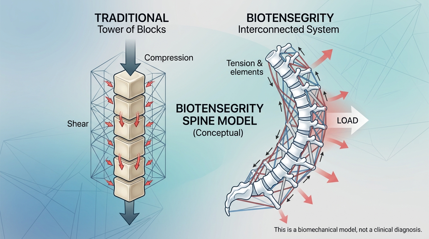
The concept has been proposed as a model for spinal mechanics and later extended to the broader bone-muscle-fascial system. Stephen Levin's influential 2002 paper proposed a tensegrity-truss model of the spine as an alternative conceptual model to the traditional “tower of blocks” representation. [1]
However, an important distinction is necessary:
Biotensegrity is a biomechanical model, not an established clinical diagnosis or a proven explanation for the cause of scoliosis.
Its value lies primarily in helping us think about the spine as a dynamic, interconnected mechanical system.
---
1. What Is Tensegrity?
Tensegrity describes a structural arrangement in which tension and compression work together to create stability.
A simple tensegrity structure may contain:
- Rigid compression elements
- Flexible tension elements
- Pre-tension
- Distributed forces
- Spatial relationships between components
Unlike a conventional stack of blocks, the components do not necessarily need to be directly connected to one another to create a stable structure.
The tension elements help maintain the position of the compression elements.
This creates a system in which changing one component can influence forces elsewhere.
A simple example
Imagine several rigid rods suspended within a network of elastic cords.
The rods do not simply sit on top of one another.
Instead, the cords hold them in position.
If one cord becomes tighter or looser, the position of several rods may change.
The entire structure responds.
This is the basic idea that makes tensegrity interesting from a biomechanical perspective.
---
2. The Traditional “Stack of Blocks” Model of the Spine
A simple representation of the spine is a stack of vertebrae separated by intervertebral discs.
This model is useful for understanding basic anatomy.
The vertebrae appear to form a column:
Vertebra → disc → vertebra → disc → vertebra
Gravity pushes downward.
The vertebrae transmit compression.
Muscles and ligaments help prevent excessive movement.
But this model becomes less satisfactory when we try to explain the spine during movement.
The spine does not simply stand vertically.
It:
- Bends
- Rotates
- Translates
- Absorbs shocks
- Responds to external loads
- Changes shape during breathing
- Adjusts during walking
- Responds to muscle activation
A purely rigid-column model therefore cannot capture the full complexity of spinal movement.
Levin argued that a tensegrity-truss model could provide a different conceptual framework for understanding spinal mechanics, particularly because forces can be distributed through tension and compression rather than being treated simply as loads passing through a rigid stack. [1]
---
3. The Spine as a Dynamic Support System
The human spine contains both relatively rigid and relatively flexible components.
Compression-bearing structures
These include:
- Vertebral bodies
- Intervertebral discs
- Facet joints
- Certain bony structures
Tension-bearing structures
These include:
- Muscles
- Tendons
- Ligaments
- Fascial tissues
- Other connective tissues
These categories are simplified because biological tissues can perform multiple mechanical functions depending on their location and loading condition.
The important point is that the spine is not stabilized by bone alone.
Muscles and connective tissues continuously influence its mechanical behavior.
This creates a useful conceptual relationship:
Bone provides structural support
*
Soft tissues regulate tension
*
Muscles actively modify the system
=
Dynamic spinal stability
---
4. Why Tension Matters
Tension is a pulling force.
Muscles generate tension when they contract.
Ligaments develop tension when joints move toward the limits of their normal range.
Tendons transmit muscle-generated forces to bones.
Fascial tissues can also transmit mechanical forces between anatomical regions.
The body therefore contains numerous interconnected tension pathways.
A change in tension in one region may influence movement or loading elsewhere.
For example:
Foot position
↓
Leg alignment
↓
Pelvic position
↓
Lumbar alignment
↓
Thoracic position
↓
Head position
This does not mean that every postural change at the foot causes a spinal deformity.
Rather, it demonstrates that the musculoskeletal system is mechanically interconnected.
---
5. Pre-Tension and Stability
One of the most important concepts in tensegrity is pre-tension.
A tensegrity structure can remain stable because tension exists throughout the system before an external load is applied.
The human body also contains baseline muscle and connective-tissue tension.
Even when standing quietly, muscles are not completely inactive.
The nervous system continuously regulates muscle activity to maintain posture.
This background activity contributes to:
- Joint stability
- Balance
- Upright posture
- Movement readiness
- Response to perturbations
The amount of muscle activation changes according to the task.
Standing quietly requires one pattern.
Walking requires another.
Lifting a heavy object requires another.
Running requires another.
Therefore, biological “tension” is not fixed.
It is continuously regulated.
---
6. Posture as a Dynamic Equilibrium
Good posture is sometimes interpreted as a perfectly symmetrical static position.
Biomechanically, this is too simple.
A living person is constantly moving, even when apparently standing still.
Small movements occur at:
- The ankles
- Knees
- Hips
- Pelvis
- Spine
- Shoulders
- Head
The nervous system continually adjusts muscle activation to maintain equilibrium.
A more useful definition of posture is therefore:
The continuously changing relationship between body segments as the neuromuscular system maintains balance and controls movement.
This fits naturally with a tensegrity-based conceptual framework.
The body does not need every component to remain motionless.
It needs the system to remain sufficiently organized while movement occurs.
---
7. Tensegrity and Spinal Alignment
Spinal alignment depends on the relationship between multiple anatomical regions.
Important components include:
- Vertebral geometry
- Intervertebral discs
- Ligaments
- Paraspinal muscles
- Abdominal muscles
- Thoracolumbar fascia
- Rib cage
- Pelvis
- Hip joints
- Lower extremities
A change in one region may alter the mechanical requirements of another.
For example, pelvic displacement may change the orientation of the lumbar spine.
Lumbar changes may influence thoracic compensation.
Thoracic changes may alter head position.
The nervous system then adjusts muscle activation to maintain balance.
This creates a continuous feedback loop:
Alignment → mechanical forces → sensory feedback → muscle activation → movement → new alignment
Tensegrity provides one way of conceptualizing this distributed mechanical interaction.
---
8. Tensegrity and the Three Dimensions of the Spine
Scoliosis is a three-dimensional deformity.
It involves:
- Coronal curvature
- Sagittal alignment
- Axial rotation
Therefore, a useful biomechanical model should also consider forces in three dimensions.
Coronal plane
The system must control:
- Lateral displacement
- Side bending
- Pelvic obliquity
Sagittal plane
The system must control:
- Flexion
- Extension
- Thoracic kyphosis
- Lumbar lordosis
- Pelvic tilt
Transverse plane
The system must control:
- Axial rotation
- Rib-cage rotation
- Pelvic rotation
A simple “straight versus curved” model does not adequately describe this complexity.
---
9. Tensegrity and Scoliosis
The tensegrity model has attracted interest in spinal biomechanics because scoliosis involves complex relationships between skeletal geometry, muscle forces, connective tissues, and movement.
However, this needs to be interpreted carefully.
There is currently no established evidence that:
“An abnormal tensegrity system causes idiopathic scoliosis.”
Idiopathic scoliosis is multifactorial, and its exact etiology remains incompletely understood.
A tensegrity model may instead help describe how mechanical forces interact after or alongside the development of spinal asymmetry.
For example:
Structural asymmetry
↓
Changed muscle length and mechanical demand
↓
Altered muscle activation
↓
Changed force distribution
↓
Postural compensation
↓
Further changes in mechanical loading
This is a conceptual feedback model rather than a proven causal pathway.
---
10. The Muscle-Fascia Network
The biotensegrity concept has also been applied to the relationship between muscles, fascia, and bones.
A 2017 article in the Italian Journal of Anatomy and Embryology described the bone-muscle-fascial system through a biotensegrity framework and discussed mechanical communication within the connective-tissue system. The article is published under a Creative Commons Attribution 4.0 license. [2]
From a clinical perspective, this concept is useful because muscles do not work as completely isolated structures.
They interact through:
- Tendons
- Aponeuroses
- Fascial connections
- Intermuscular tissues
- Joint structures
- Neural control
However, the existence of mechanical connections does not mean that every fascial pathway functions as a single continuous cable capable of transmitting unlimited force.
Biological tissues are complex, viscoelastic, nonlinear, and regionally specialized.
Therefore, biotensegrity should be used as a conceptual framework rather than a literal engineering diagram of the human body.
---
11. Core Stability as an Active Tension System
Core stability can be interpreted within this model.
The trunk contains multiple muscles that continuously regulate spinal position.
These include:
- Multifidus
- Erector spinae
- Transversus abdominis
- Internal oblique
- External oblique
- Rectus abdominis
- Quadratus lumborum
- Diaphragm
- Pelvic floor muscles
Their roles vary according to posture and movement.
Instead of thinking:
“The core holds the spine rigid.”
A more accurate model is:
The core continuously modulates spinal stiffness and movement according to the mechanical demands of the task.
This distinction is particularly important for dynamic posture.
---
12. The Spine Does Not Need Maximum Tension
More tension is not always better.
Excessive muscle activation may increase:
- Compression
- Energy expenditure
- Fatigue
- Stiffness
Insufficient activation may reduce:
- Stability
- Movement control
- Resistance to external forces
The nervous system therefore attempts to find an appropriate level of activation.
This can be described as:
Minimum necessary stiffness + sufficient control + appropriate mobility
The ideal posture is consequently not necessarily the posture with the greatest muscular tension.
It is the posture in which the body can perform the required task efficiently and safely.
---
13. Dynamic Versus Rigid Support
This concept connects directly with the difference between dynamic tension and rigid fixation.
Rigid support
Rigid fixation attempts to control spinal position through:
- Screws
- Rods
- Fusion
- Structural implants
The mechanical relationship between selected spinal segments becomes substantially constrained.
Dynamic support
Dynamic systems attempt to preserve some movement while influencing force distribution.
Examples may include:
- Flexible braces
- Elastic support systems
- Exercise-based stabilization
- Active self-correction
- Selected growth-modulation techniques
These approaches operate through different mechanisms and should not be considered interchangeable.
Tensegrity is particularly relevant to the conceptual idea of dynamic support, because stability can emerge from continuously interacting forces rather than from immobilization.
---
14. Tensegrity and Movement
One of the most interesting features of a tensegrity-inspired model is that changing one component can influence the entire structure.
The same principle can be used conceptually to understand movement.
When the arm moves forward:
Arm movement
↓
Shoulder girdle
↓
Thorax
↓
Spinal muscles
↓
Pelvis
↓
Lower extremities
The body does not need to consciously control every individual segment.
The nervous system coordinates the entire movement.
This is why posture and movement should not always be separated.
A person may have excellent static alignment but poor dynamic control.
Another person may appear asymmetrical while standing but move efficiently during walking.
---
15. Tensegrity and Neuromuscular Feedback
Neuromuscular feedback provides another layer to the model.
The body continuously receives information from:
- Muscles
- Tendons
- Joints
- Skin
- Vestibular system
- Vision
The nervous system uses these signals to modify muscle activation.
The cycle is:
Mechanical position
↓
Sensory information
↓
Central processing
↓
Motor response
↓
Changed force distribution
↓
New mechanical position
This means that posture is not simply a mechanical problem.
It is also a sensorimotor problem.
A tensegrity-based model describes the mechanical network, while neuromuscular feedback describes how the nervous system continuously regulates that network.
---
16. Tensegrity and Pelvic Alignment
The pelvis is an important part of the body's mechanical network.
It connects the spine with the lower extremities.
Changes in pelvic position can influence:
- Lumbar lordosis
- Trunk position
- Hip mechanics
- Weight distribution
- Standing balance
Potential contributors to pelvic asymmetry include:
- Leg-length discrepancy
- Hip mobility
- Muscle imbalance
- Foot position
- Structural scoliosis
- Functional compensation
A tensegrity-based framework encourages clinicians to consider these relationships rather than treating the spine as an isolated structure.
However, pelvic asymmetry does not automatically mean that the pelvis is the original cause of scoliosis.
The direction of causation must be determined from the clinical context.
---
17. Tensegrity and the Rib Cage
The rib cage is another important component.
The thoracic spine and ribs form a mechanically integrated structure.
In scoliosis, vertebral rotation may produce changes in rib position and trunk shape.
The resulting asymmetry may influence:
- Breathing mechanics
- Muscle activation
- Trunk rotation
- Postural control
- Perception of body position
This is one reason three-dimensional scoliosis rehabilitation may incorporate breathing and rib-cage movement.
The objective is not simply to make the ribs “straight.”
It is to improve the patient's ability to control thoracic movement and breathing within the existing structural anatomy.
---
18. What Happens When Force Distribution Becomes Asymmetric?
An asymmetric spine does not necessarily mean that every force becomes harmful.
The body can compensate.
For example, one region may experience greater muscular demand while another region contributes less.
Over time, however, a persistent asymmetry may influence:
- Muscle endurance
- Movement efficiency
- Fatigue
- Joint loading
- Postural strategy
This creates an important clinical distinction:
Asymmetry is not automatically pathology.
The human body is naturally asymmetrical.
The relevant question is whether the asymmetry is associated with:
- Structural progression
- Pain
- Functional limitation
- Reduced balance
- Excessive fatigue
- Neurological findings
- Impaired movement
---
19. Tensegrity Is Not the Same as “Fascia Causes Everything”
Tensegrity and fascia are frequently discussed together online.
This can lead to an oversimplified claim:
“Fascial tension causes spinal deformity.”
Current evidence does not justify such a universal statement.
Fascia certainly participates in mechanical force transmission, but scoliosis is a complex condition involving skeletal development, genetics, neuromuscular factors, growth, biomechanics, and other biological processes.
Therefore, it is more accurate to say:
Fascial and muscular tissues contribute to the mechanical environment of the spine and may influence movement and load distribution.
That is substantially different from claiming that fascial tension is the primary cause of scoliosis.
---
20. Tensegrity and Exercise-Based Postural Correction
Tensegrity concepts can provide a useful framework for understanding why exercise-based correction may involve more than strengthening individual muscles.
A patient may be trained to coordinate:
- Trunk elongation
- Pelvic control
- Rib-cage positioning
- Rotation control
- Breathing
- Muscle endurance
- Balance
The objective is to modify the whole movement strategy.
This is consistent with the broader principle of scoliosis-specific exercise, in which the patient's individual curve pattern and three-dimensional alignment are considered.
However, the clinical effectiveness of any exercise approach should be evaluated using appropriate outcomes rather than assumed from a theoretical tensegrity model.
---
21. A Patient-Specific Tensegrity Perspective
Every patient's mechanical system is different.
Relevant variables include:
- Curve type
- Cobb angle
- Vertebral rotation
- Sagittal alignment
- Pelvic position
- Skeletal maturity
- Muscle characteristics
- Balance
- Flexibility
- Pain
- Activity level
Therefore, a single “ideal” tension pattern cannot be assumed for every patient.
One patient may require greater control of trunk rotation.
Another may need improved pelvic stability.
Another may have a major issue with endurance.
Another may have significant structural rigidity that cannot be substantially changed through exercise.
A useful tensegrity-based assessment therefore asks:
Where is the load?
Where is the tension?
Where is the compression?
Where does the body compensate?
Which muscles are actively controlling the position?
Can the patient change the alignment voluntarily?
Does the correction remain stable during movement?
---
22. Computational Models of Spinal Tensegrity
Researchers have constructed computational and physical models inspired by the tensegrity concept.
These models can investigate:
- Spinal deformation
- Force transmission
- Axial loading
- Shape change
- Mechanical stability
- Dynamic movement
Open-access engineering studies have developed spine-inspired biotensegrity models that reproduce aspects of natural spinal curvature and investigate multidirectional shape change. [3][4]
These models are useful because they allow researchers to manipulate individual parameters that cannot easily be isolated in living humans.
For example, a model can change:
- Cable tension
- Compression stiffness
- Segment geometry
- External loading
- Direction of applied force
Researchers can then observe how the overall structure responds.
However, a computational model remains a model.
It does not prove that the human spine operates identically to the engineered system.
---
23. Tensegrity and Future Personalized Modeling
Future scoliosis research may combine tensegrity-inspired models with patient-specific data.
Potential inputs could include:
- Standing X-rays
- EOS imaging
- CT or MRI
- Motion capture
- Surface EMG
- Force plates
- Wearable sensors
- Muscle morphology
- Skeletal maturity
A computational model could potentially estimate how changes in:
- Muscle activation
- External support
- Postural strategy
- Loading direction
might influence a particular patient's spinal mechanics.
This could eventually contribute to more individualized rehabilitation and orthotic design.
At present, however, these applications remain primarily research-oriented.
---
24. The Clinical Meaning of Tensegrity-Based Posture Alignment
For patients, the practical message is relatively simple.
The spine should not be thought of as a rigid tower that simply needs to be pushed into a straight position.
It is a living system in which:
- Bones provide structural support.
- Discs permit controlled movement.
- Ligaments constrain excessive motion.
- Muscles actively regulate stability.
- Connective tissues transmit mechanical forces.
- The nervous system continuously adjusts the system.
Tensegrity provides one conceptual language for describing this interaction.
It encourages attention to relationships rather than isolated structures.
Instead of asking only:
“Where is the curve?”
we can also ask:
“How does the rest of the body respond to the curve?”
---
25. Tensegrity and the Goal of Postural Alignment
The goal of postural alignment should not necessarily be perfect geometric symmetry.
Functional alignment is more important.
A well-controlled posture allows the individual to:
- Stand comfortably
- Walk efficiently
- Move through different positions
- Maintain balance
- Control external loads
- Breathe effectively
- Avoid unnecessary muscular effort
In this context, alignment is dynamic.
The body should be capable of changing position while maintaining sufficient stability.
This is perhaps the most useful connection between tensegrity and posture.
Stability does not require immobility.
---
26. A Conceptual Model for Scoliosis
A simplified tensegrity-inspired model of scoliosis can be represented as:
Structural asymmetry
↓
Changed geometry
↓
Changed muscle length and mechanical demand
↓
Changed neuromuscular feedback
↓
Changed muscle activation
↓
Changed force distribution
↓
Postural compensation
↓
New mechanical environment
This model should not be interpreted as a proven mechanism of idiopathic scoliosis progression.
Rather, it illustrates how structural and functional factors may interact once spinal asymmetry exists.
---
27. JESU5C and Pattern-Based Mechanical Assessment
A pattern-based approach to scoliosis may attempt to classify individual patients according to the distribution of curvature, rotation, posture, muscle tension, and mechanical loading.
The JESU5C pattern classification, Meridian-Based Isometric Exercises, and HGMT represent a clinician-developed framework that approaches scoliosis through individual structural and functional patterns. Such a framework can be discussed alongside biomechanical concepts such as dynamic tension and force distribution.
However, it should be distinguished clearly from established radiographic classifications and validated clinical systems.
Its specific diagnostic or therapeutic claims require independent clinical research before they can be considered established evidence.
The potential value of such a framework lies in generating testable hypotheses about how different alignment patterns may respond to individualized mechanical strategies.
---
28. What Tensegrity Can—and Cannot—Explain
Tensegrity can help conceptualize:
- Distributed mechanical forces
- Interaction between tension and compression
- Dynamic stability
- Muscle-generated forces
- Whole-body mechanical relationships
- Movement-dependent changes in alignment
- Postural adaptation
Tensegrity cannot by itself establish:
- The cause of idiopathic scoliosis
- A specific treatment for scoliosis
- That fascia is the primary driver of spinal deformity
- That correcting muscle tension will straighten a structural curve
- That every postural asymmetry represents pathological tension imbalance
This distinction protects the concept from becoming a metaphor that is presented as established clinical fact.
---
Conclusion
Tensegrity offers a useful way to think about posture and spinal alignment as a dynamic system of interacting forces rather than a rigid stack of bones.
The spine exists within a network involving:
bone + discs + ligaments + muscles + fascia + pelvis + rib cage + nervous system.
Changes in one part of this network can alter mechanical demands elsewhere.
For scoliosis, this perspective may help explain why patients with similar Cobb angles can have different postural strategies, muscle activation patterns, symptoms, and functional abilities.
However, tensegrity remains primarily a conceptual biomechanical model. It should not be presented as a proven cause of scoliosis or as evidence that fascial or muscular tension alone can correct structural deformity.
The most useful interpretation is more balanced:
The spine is a dynamic structure whose alignment and stability emerge from the interaction of skeletal geometry, passive tissues, active muscular forces, and neuromuscular control.
Understanding these relationships may eventually contribute to more individualized approaches to posture assessment, exercise, bracing, and spinal biomechanics.
For patients with scoliosis, the ultimate objective is not simply to create a perfectly straight spine.
It is to achieve an appropriate balance between:
stability + mobility + force distribution + neuromuscular control + functional movement.
That is where the tensegrity concept may have its greatest value: not as a replacement for established scoliosis science, but as a framework for asking better questions about how the whole musculoskeletal system supports and adapts to spinal alignment.
References
[1] Levin, S. M. “The Tensegrity-Truss as a Model for Spine Mechanics: Biotensegrity.” Journal of Mechanics in Medicine and Biology, 2002. DOI.
[2] Saggini, R. “Posture as a model of mechanosensitivity: the ‘Biotensegrity’.” Italian Journal of Anatomy and Embryology, 2017. Original article.
[3] Oh, C. L., Choong, K. K., Nishimura, T., & Kim, J.-Y. “Form-Finding of Spine Inspired Biotensegrity Model.” Applied Sciences, 2020, 10(18), 6344. Original article.
[4] Oh, C. L., Choong, K. K., Nishimura, T., & Kim, J.-Y. “Multi-Directional Shape Change Analysis of Biotensegrity Model Mimicking Human Spine Curvature.” Applied Sciences, 2022, 12(5), 2377. Original article.
[5] Bordoni, B., Marelli, F., Morabito, B., & Castagna, R. “A New Concept of Biotensegrity Incorporating Liquid Tissues: Blood and Lymph.” Journal of Evidence-Based Integrative Medicine, 2018. Original article.
[6] LEE, Yongsub, & Lee, Minsu (translator). Scoliosis, Meridian-Based Isometric Exercises and JESU5C Pattern Suit: “JESU5C” a New Classification of Idiopathic Scoliosis and HGMT (Hyun-Ga Manual Technique). Amazon Kindle Edition.