Spine Care
[Biomechanics, Tensegrity & Meridian Concepts 10] The Connection Between Spinal Alignment and Neuromuscular Feedback
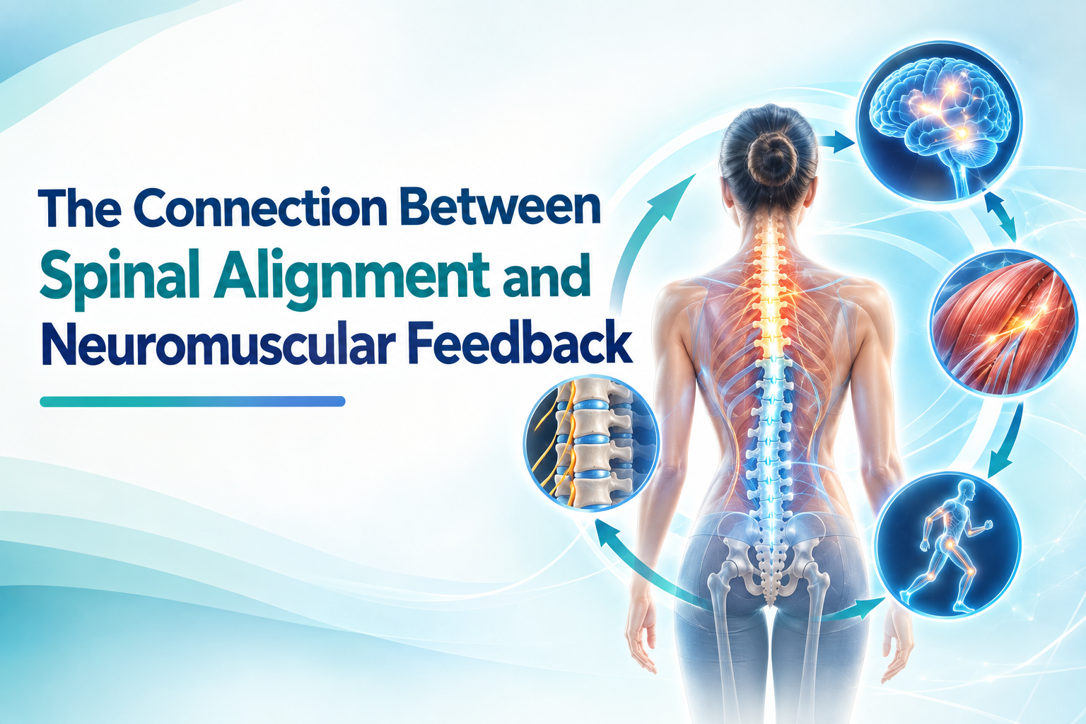
Spinal alignment and neuromuscular feedback constantly interact through proprioception, muscle activation, balance, and postural control.
Introduction
The spine is not a passive column that simply responds to gravity. It is part of a continuously regulated neuromuscular system. Every time a person stands, walks, bends, reaches, or changes direction, the nervous system receives information about body position and adjusts muscle activity to maintain balance and movement.
This ongoing process is known as neuromuscular feedback.
For people with scoliosis, this relationship is particularly important because spinal alignment is three-dimensional. Changes in coronal alignment, sagittal alignment, vertebral rotation, pelvic position, and trunk symmetry can alter the mechanical information available to the nervous system. In turn, altered sensory information and muscle activation may influence how a person maintains posture and moves.
Research has identified differences in proprioception, postural stability, muscle activation, and movement patterns in adolescents with idiopathic scoliosis (AIS). However, these findings do not establish that abnormal neuromuscular feedback causes idiopathic scoliosis. The relationship is more likely to be complex and bidirectional, with spinal structure and neuromuscular control influencing one another over time. [1][2][3]
Understanding this connection helps explain why evaluating scoliosis involves more than measuring the Cobb angle on a standing X-ray.
---
1. What Is Neuromuscular Feedback?
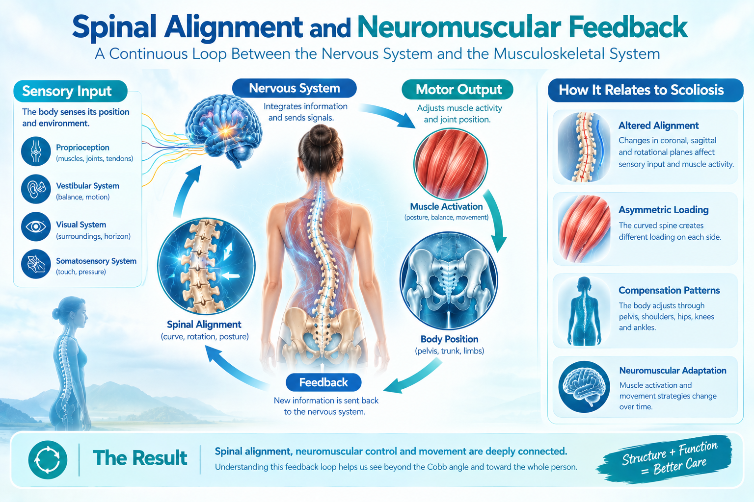
Neuromuscular feedback is the continuous exchange of information between the nervous system and the musculoskeletal system.
The nervous system receives information about:
- Joint position
- Muscle length
- Muscle tension
- Body orientation
- Movement speed
- Ground contact
- Balance
- Head position
- Trunk position
- Visual surroundings
It then uses this information to regulate muscle activity.
For example, when a person begins to lean toward the right, the nervous system detects the change through sensory systems and produces corrective muscle activation. The person may shift the pelvis, activate trunk muscles, move the arms, or adjust the feet before consciously recognizing the imbalance.
This process happens continuously during standing and movement.
The major sensory systems
Postural control depends on the integration of information from several systems:
Proprioceptive system: Information from muscles, tendons, joints, and other tissues about body position and movement.
Vestibular system: Information about head movement, acceleration, and orientation relative to gravity.
Visual system: Information about the body's relationship to the surrounding environment.
Somatosensory system: Information about pressure, touch, and contact with surfaces.
The brain integrates these signals and generates appropriate motor responses.
---
2. Proprioception: The Body's Internal Position Sensor
Proprioception is particularly relevant to spinal alignment.
A person does not need to look at their spine to know that they are leaning. Receptors throughout the musculoskeletal system provide information that allows the nervous system to estimate body position.
This information contributes to:
- Postural control
- Joint stability
- Movement coordination
- Balance
- Muscle activation
- Spatial orientation
Research suggests that adolescents with idiopathic scoliosis may have differences in proprioceptive function compared with individuals without scoliosis.
A systematic review and meta-analysis involving 11 case-control studies and 1,121 participants found evidence of proprioceptive deficits in AIS, including greater repositioning errors and higher motion-detection thresholds. However, the authors emphasized that the causal relationship between scoliosis and proprioceptive deficits remains uncertain. [1]
This distinction is important.
A proprioceptive difference may:
- contribute to altered postural control,
- develop as an adaptation to an altered spinal structure,
- coexist with another neurological or biomechanical factor, or
- reflect a combination of these mechanisms.
Therefore, proprioceptive impairment should not be interpreted as a proven cause of idiopathic scoliosis.
---
3. How the Spine Provides Feedback to the Nervous System
The spine contains numerous structures capable of providing sensory information.
These include:
- Facet joints
- Intervertebral discs
- Ligaments
- Paraspinal muscles
- Tendons
- Skin and superficial tissues
- Other mechanosensitive structures
When spinal alignment changes, the mechanical environment of these tissues also changes.
A rotated vertebra may alter muscle length on one side and compression or tension on another. A pelvic shift can change the relative position of the trunk over the lower extremities. A sagittal imbalance can modify the amount of muscular effort required to maintain an upright position.
The nervous system receives information from these changing mechanical conditions.
This creates an important feedback loop:
Spinal position → sensory information → central processing → muscle activation → movement/posture → new spinal position
The loop operates continuously.
---
4. Muscle Activation Is Part of the Feedback System
The paraspinal muscles are not simply structures that “hold the spine up.” They actively participate in postural regulation.
When the trunk moves, muscles can increase or decrease their activation depending on:
- Direction of movement
- Spinal position
- External load
- Balance requirements
- Expected movement
- Fatigue
- Pain
- Previous movement experience
Electromyography (EMG) allows researchers to study this activity.
Research on AIS has found evidence of asymmetrical paraspinal muscle activity in some situations, but the findings are not completely consistent.
A 2022 narrative review analyzed 136 EMG outcome measures from 16 studies. Forty-three measures supported greater convex-side activation, while 85 showed no significant side-to-side difference and eight favored greater concave-side activation. Convex-side activity appeared more frequently at the curve apex, particularly during postural tasks and in some patients with progressive right thoracic curves. [2]
This variability is clinically important.
It means that the statement “the convex-side muscles are always tighter or stronger” is too simplistic.
Muscle activation can change according to:
- Curve location
- Curve magnitude
- Task
- Body position
- Muscle examined
- Stage of progression
- Individual motor strategy
---
5. The Concave and Convex Sides May Behave Differently
Structural scoliosis creates asymmetric mechanical conditions.
On the concave side, tissues may experience relative shortening or compression. On the convex side, tissues may experience relative lengthening and altered mechanical demand.
However, muscle behavior does not simply follow this geometric pattern.
A systematic review of paraspinal muscle characteristics in conservatively treated AIS found low- to very-low-strength evidence for several differences between the two sides. The convex side was associated in some studies with greater muscle volume, more type I fibers, and greater muscle activity, whereas the concave side showed greater fatty infiltration and other signs of altered muscle characteristics. The authors emphasized that these changes may be either causes or consequences of the spinal curvature. [4]
This supports a more sophisticated model:
Spinal deformity ↔ altered muscle structure ↔ altered muscle activation ↔ altered loading
Rather than assuming that one side simply becomes “weak” and the other “tight,” clinicians should consider how each muscle participates in the patient's specific movement strategy.
---
6. Postural Control Depends on Continuous Feedback
Standing still appears simple, but it is actually a dynamic process.
The body constantly makes small adjustments to keep the center of mass within an appropriate relationship to the base of support.
These adjustments are controlled through:
- Ankle muscles
- Hip muscles
- Pelvic muscles
- Trunk muscles
- Paraspinal muscles
- Vestibular information
- Proprioceptive information
- Vision
A systematic review and meta-analysis found moderate-quality evidence that adolescents with idiopathic scoliosis have differences in postural stability, including increased sway-area and changes in mediolateral and anteroposterior center-of-pressure parameters. [3]
A more recent systematic review integrating neurophysiological, balance, and motion research also found evidence of impaired standing balance in AIS, along with differences in muscle activation and other neurophysiological measures. [5]
These findings suggest that scoliosis can involve not only an altered spinal shape but also an altered postural-control strategy.
---
7. From Static Alignment to Dynamic Alignment
A standing X-ray provides an important picture of spinal structure, but it captures only one moment.
Human movement is dynamic.
During walking, for example:
- The pelvis rotates.
- The trunk moves relative to the pelvis.
- The arms swing.
- The spine changes its orientation.
- Muscles repeatedly change activation.
- Ground-reaction forces change.
- Balance corrections occur continuously.
A 2026 review of reviews found consistent associations between AIS and differences in gait, pelvic and trunk motion, balance, postural control, muscle activation, loading patterns, and spinal mobility. The authors emphasized that static radiographic measurements do not fully capture how scoliosis is expressed during movement. [6]
This is an important concept for understanding neuromuscular feedback.
Two patients may have similar Cobb angles but demonstrate very different movement strategies.
One may maintain relatively stable trunk control during walking, while another may rely heavily on compensatory pelvic or shoulder movements.
---
8. The Feedback Loop Can Become a Learned Movement Strategy
Repeated movement creates motor learning.
If the body repeatedly encounters the same alignment challenge, the nervous system may develop a familiar strategy for dealing with it.
For example:
Initial asymmetry
↓
The body detects altered balance
↓
Muscles increase activity to stabilize the trunk
↓
The pelvis or shoulders adjust
↓
The altered position becomes repeatedly practiced
↓
The nervous system becomes accustomed to the strategy
↓
The movement pattern becomes increasingly automatic
This does not necessarily mean that the nervous system is “causing” the scoliosis.
Rather, the nervous system may learn to operate efficiently within the altered mechanical environment.
This distinction is particularly important in chronic conditions.
A movement strategy that initially served as compensation may eventually become habitual.
---
9. Compensation Can Be Helpful
Not every asymmetry is harmful.
Compensation is often necessary for maintaining function.
For example, if the pelvis shifts slightly to one side, the trunk may move in the opposite direction to keep the head approximately centered over the feet.
This allows the person to:
- Maintain balance
- Keep the eyes level
- Continue walking
- Perform daily activities
- Reduce immediate instability
The body therefore does not necessarily seek perfect symmetry.
Instead, the nervous system seeks an acceptable combination of:
stability + efficiency + movement + sensory accuracy
A postural asymmetry may therefore represent an adaptive strategy rather than a primary structural abnormality.
---
10. When Compensation Becomes Less Efficient
A compensation strategy can become problematic when it requires excessive muscular effort or becomes mechanically inefficient.
Possible contributing factors include:
- Increasing curve magnitude
- Severe vertebral rotation
- Pelvic obliquity
- Leg-length discrepancy
- Muscle fatigue
- Pain
- Reduced mobility
- Poor balance
- Limited proprioceptive accuracy
- Reduced physical conditioning
As the mechanical demands increase, the nervous system may need to increase muscle activation to maintain the same posture.
This can contribute to fatigue and discomfort.
The relationship can therefore become cyclical:
Altered alignment → increased stabilization demand → altered muscle activation → fatigue or discomfort → further movement adaptation
This is one reason why a patient may report that standing for long periods becomes increasingly uncomfortable even when the radiographic curve has not changed dramatically.
---
11. Neuromuscular Feedback and Pain
Pain can significantly change motor control.
When a region becomes painful, the nervous system may alter muscle activation to protect it.
This may include:
- Increased co-contraction
- Reduced movement
- Muscle guarding
- Changes in movement speed
- Altered load distribution
- Avoidance of particular positions
Therefore, muscle tightness does not necessarily mean that a muscle imbalance caused the spinal deformity.
A muscle may become tight because it is repeatedly working to stabilize an already asymmetric structure.
This is another example of a bidirectional relationship between structure and neuromuscular control.
---
12. The Role of Muscle Co-Contraction
Co-contraction occurs when muscles on opposing sides of a joint or body segment activate simultaneously.
It can increase stability, particularly when the nervous system perceives uncertainty or instability.
In scoliosis, increased co-contraction may help maintain upright posture despite altered spinal geometry.
However, excessive co-contraction can also increase energy expenditure and muscle fatigue.
Research examining postural control in AIS suggests that individuals may use altered muscle activation strategies to maintain balance. [3][5]
Therefore, a patient's ability to remain upright does not necessarily mean that their postural system is functioning normally.
The body may be maintaining stability through a more demanding strategy.
---
13. Visual Feedback and Spinal Alignment
Vision provides another important source of postural information.
When standing, the brain continuously compares the position of the body with the surrounding environment.
For example, visual information can help a person recognize:
- Whether the head is tilted
- Whether the shoulders are level
- Whether the body is leaning
- Whether the pelvis appears shifted
- Whether the horizon is level
When visual information is removed, the nervous system must rely more heavily on proprioceptive and vestibular information.
Studies of AIS have investigated how individuals use these sensory systems during balance tasks. Evidence suggests that visual and proprioceptive information can play important roles in maintaining postural stability in AIS. [5]
This helps explain why exercises performed with mirrors or visual feedback may sometimes help patients learn a new postural strategy.
---
14. Why Mirror Feedback Can Be Useful
Visual feedback can make an otherwise invisible alignment problem visible to the patient.
A patient may be asked to observe:
- Shoulder level
- Pelvic position
- Trunk shift
- Rib prominence
- Head position
- Weight distribution
The patient can then attempt an active correction while watching the change.
This creates a new feedback loop:
See → correct → feel → repeat → learn
Over time, repeated practice may help the patient develop better awareness and control of the desired position.
This principle is consistent with the broader concept of active self-correction used in scoliosis-specific rehabilitation.
However, mirror-based correction should not be interpreted as proof that a structural curve can simply be “straightened” through conscious posture. Structural deformity and functional posture are different phenomena.
---
15. Exercise Can Train the Neuromuscular System
Scoliosis-specific exercise approaches attempt to influence more than muscle strength.
They may address:
- Three-dimensional posture
- Active self-correction
- Trunk stabilization
- Breathing
- Balance
- Proprioception
- Movement awareness
- Muscle endurance
A review of Schroth-based interventions reported positive effects on postural control and balance, although the authors emphasized the need for further investigation. [7]
A 2026 umbrella review of exercise interventions for AIS also reported improvements in outcomes such as posture, spinal curvature measures, physical function, and quality of life across the available systematic-review evidence, while emphasizing differences among interventions and study quality. [8]
The goal of exercise should therefore not be reduced to “strengthening the weak side.”
The more sophisticated objective is to improve the patient's ability to sense, control, and repeatedly reproduce an efficient three-dimensional posture during movement.
---
16. Neuromuscular Feedback and Bracing
Bracing also changes the sensory and mechanical environment of the trunk.
A brace applies external pressure and changes how the patient experiences trunk position.
This may influence:
- Skin pressure
- Proprioceptive input
- Muscle activation
- Postural strategy
- Balance
- Movement
Research examining orthotic interventions has found that AIS patients may use altered postural-control strategies involving muscle co-contraction and proprioceptive information. However, evidence regarding the longer-term effects of bracing on balance remains limited and heterogeneous. [9]
This means that a brace should not be viewed only as a mechanical device.
It also becomes part of the patient's sensory environment.
---
17. The Spine and Nervous System as a Closed-Loop System
A useful conceptual model is to consider spinal alignment and neuromuscular control as a closed-loop system.
Structural component
The spine provides the mechanical framework:
- Vertebrae
- Discs
- Facet joints
- Ligaments
- Pelvis
- Rib cage
Sensory component
The body detects:
- Position
- Motion
- Pressure
- Muscle length
- Muscle tension
- Balance
Processing component
The nervous system integrates:
- Proprioception
- Vision
- Vestibular information
- Somatosensory information
- Previous movement experience
Motor component
The nervous system controls:
- Muscle activation
- Joint movement
- Trunk position
- Pelvic movement
- Balance reactions
Feedback component
The resulting movement changes spinal and body position, producing new sensory information.
The loop then repeats.
This framework helps explain why spinal alignment cannot always be understood from a static image alone.
---
18. What This Means for Scoliosis Assessment
A comprehensive assessment may need to consider both structure and function.
Structural assessment
Important variables include:
- Cobb angle
- Curve pattern
- Vertebral rotation
- Vertebral wedging
- Sagittal alignment
- Pelvic parameters
- Skeletal maturity
- Curve flexibility
Functional assessment
Additional information may include:
- Standing balance
- Gait
- Trunk movement
- Pelvic movement
- Muscle activation
- Postural control
- Proprioception
- Breathing mechanics
- Functional limitations
This distinction is especially useful when two patients have similar radiographic curves but very different symptoms and movement patterns.
---
19. Why Cobb Angle Alone Cannot Describe Neuromuscular Function
The Cobb angle is essential for quantifying scoliosis, but it is a structural measurement.
It does not directly measure:
- Muscle activation
- Proprioception
- Balance
- Motor learning
- Gait
- Dynamic trunk control
- Neurological processing
A 30-degree curve does not automatically tell us how efficiently a patient controls their trunk during walking.
Likewise, a small change in Cobb angle does not necessarily capture every clinically meaningful change in function.
For this reason, radiographic and functional assessments should be viewed as complementary rather than competing approaches.
---
20. A Pattern-Based Approach to Neuromuscular Feedback
A patient-specific approach can ask several questions:
Where is the structural curve?
Where is the trunk shifted?
How is the pelvis positioned?
Which muscles demonstrate altered activation?
How does the patient maintain balance?
What happens during movement rather than only standing?
Does the patient actively correct the alignment when given feedback?
Can the corrected position be maintained without excessive muscular effort?
These questions may reveal that two patients with similar Cobb angles have different neuromuscular strategies.
A clinician-developed framework such as the JESU5C pattern classification, Meridian-Based Isometric Exercises, and HGMT may be used as one conceptual approach to examining these relationships. Such a framework should be distinguished from established diagnostic classifications, and its clinical claims require independent validation through appropriately designed research. [10]
---
21. From Passive Correction to Active Control
Traditional correction strategies often emphasize externally applied forces.
Neuromuscular rehabilitation adds another dimension:
Can the patient actively control the corrected position?
This is important because a posture maintained only through an external force may not automatically become a patient's habitual movement pattern.
Active control involves:
- Detecting the current position
- Understanding the desired correction
- Producing the appropriate muscular response
- Maintaining the correction
- Repeating it during different movements
- Gradually making the response more automatic
This is essentially a process of sensorimotor learning.
The objective is not necessarily to maintain a rigid posture all day. Rather, it is to improve the ability to move through a range of positions while maintaining appropriate control.
---
22. The Importance of Movement, Not Just Position
The nervous system does not control a static spine alone.
It controls movement.
Therefore, an effective neuromuscular assessment may examine the patient while:
- Walking
- Turning
- Sitting
- Standing
- Reaching
- Bending
- Rotating
- Climbing stairs
- Performing sport-specific movements
Recent biomechanical research increasingly emphasizes movement-related differences in AIS, including gait, trunk motion, pelvic motion, muscle activation, loading, and spinal mobility. [6]
This shift from static alignment toward dynamic alignment may become increasingly important in future scoliosis assessment.
---
23. Can Neuromuscular Feedback Change the Structural Curve?
This question requires caution.
Improving neuromuscular control can potentially improve:
- Posture
- Balance
- Movement efficiency
- Muscle endurance
- Functional symmetry
- Quality of life
Some exercise interventions have also demonstrated reductions in Cobb angle or other measures of spinal deformity.
However, this does not mean that every structural scoliosis can be corrected simply by retraining the nervous system.
Structural scoliosis involves vertebral and spinal changes that may persist even when posture and motor control improve.
Therefore:
Improved neuromuscular control ≠ guaranteed structural correction.
The strongest interpretation is that neuromuscular training may influence functional alignment and may contribute to better management of scoliosis, while the magnitude of structural change depends on the patient's age, skeletal maturity, curve characteristics, treatment, and underlying biology.
---
24. A Bidirectional Model of Scoliosis
The relationship can be summarized as a bidirectional model:
Spinal structure
↓
Changes mechanical alignment
↓
Changes sensory input
↓
Changes neuromuscular response
↓
Changes muscle activation and movement
↓
Changes force distribution
↓
May influence functional alignment
↓
Provides new sensory input
↓
Feedback continues
This model does not claim that neuromuscular feedback causes idiopathic scoliosis.
Instead, it provides a framework for understanding how a pre-existing structural deformity and the nervous system may interact over time.
---
Conclusion
Spinal alignment and neuromuscular feedback are closely connected.
The spine continuously provides mechanical and sensory information to the nervous system, while the nervous system continuously regulates muscle activation, balance, posture, and movement in response. Research in adolescent idiopathic scoliosis has identified differences in proprioception, postural stability, muscle activation, and movement biomechanics, although the causal relationships remain incompletely understood. [1][2][3][5][6]
This evidence supports a broader view of scoliosis assessment.
A standing X-ray tells us what the spinal structure looks like.
Neuromuscular assessment helps us understand how the person controls and moves that structure.
The most useful clinical perspective may therefore combine:
structure + sensory feedback + muscle activation + movement + force distribution.
For patients, this means that scoliosis management should not necessarily focus only on making the spine appear straighter. It should also consider whether the individual can maintain an efficient, stable, and adaptable movement strategy.
Future scoliosis research may increasingly combine three-dimensional imaging, surface EMG, motion capture, force-plate analysis, wearable sensors, and computational modeling to understand how spinal structure and neuromuscular control interact in individual patients.
The ultimate goal is not simply to identify where the spine is curved, but to understand how the entire neuromusculoskeletal system responds to that curvature—and how that response might be safely improved.
References
[1] Yao, G., et al. “Timely Revisit of Proprioceptive Deficits in Adolescent Idiopathic Scoliosis: A Systematic Review and Meta-Analysis.” PubMed. 2022.
[2] Ng, P. T. T., Claus, A., Izatt, M. T., Pivonka, P., & Tucker, K. “Is spinal neuromuscular function asymmetrical in adolescents with idiopathic scoliosis compared to those without scoliosis?: A narrative review of surface EMG studies.” PubMed. Journal of Electromyography and Kinesiology, 2022.
[3] Dufvenberg, M., Adeyemi, F., Rajendran, I., Öberg, B., & Abbott, A. “Does postural stability differ between adolescents with idiopathic scoliosis and typically developed? A systematic literature review and meta-analysis.” PubMed. Scoliosis and Spinal Disorders, 2018.
[4] “Associations between paraspinal muscle characteristics and spinal curvature in conservatively treated adolescent idiopathic scoliosis: a systematic review.” PubMed. 2023.
[5] “Neurophysiological, balance and motion evidence in adolescent idiopathic scoliosis: A systematic review.” PubMed. 2024.
[6] Chockalingam, N., et al. “Movement Related Biomechanics in Adolescent Idiopathic Scoliosis: A Review of Reviews.” PubMed. 2026.
[7] “The Effect of Schroth Method on Postural Control and Balance in Patients with Adolescent Idiopathic Scoliosis: A Literature Review.” PubMed. 2023.
[8] “Effectiveness of exercise interventions for adolescent idiopathic scoliosis: An umbrella review.” PubMed. 2026.
[9] “The effect of orthotic interventions on balance performance in adolescent idiopathic scoliosis: A systematic literature review.” PubMed. 2019.
[10] LEE, Yongsub, & Lee, Minsu (translator). Scoliosis, Meridian-Based Isometric Exercises and JESU5C Pattern Suit: “JESU5C” a New Classification of Idiopathic Scoliosis and HGMT (Hyun-Ga Manual Technique). Amazon Kindle Edition.