Spine Care
[Biomechanics, Tensegrity & Meridian Concepts 05] Asymmetric Muscle Tone: Why One Side of Your Back Is Tighter
Learn how muscle tone, stiffness, spinal rotation, posture, and asymmetric loading interact—and what the research says about scoliosis and muscle imbalance.
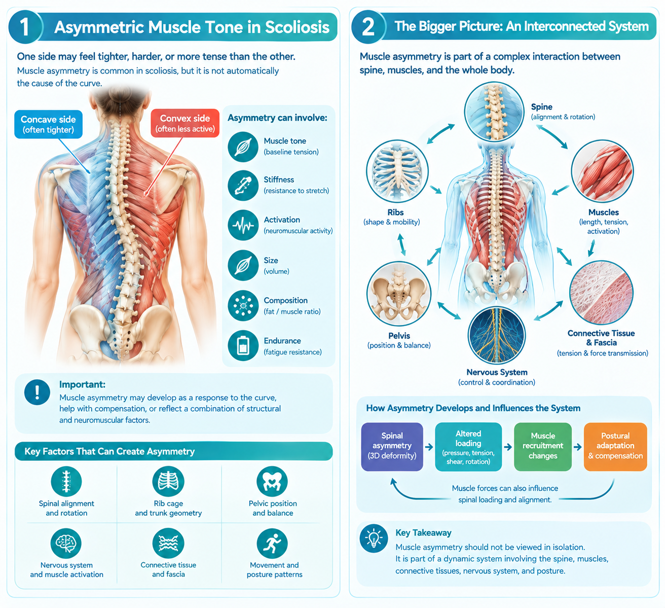
If one side of your back feels tighter, harder, or more tense than the other, you may naturally wonder whether the difference is caused by scoliosis. In many people with scoliosis, the muscles on the two sides of the spine do not behave identically. Differences can occur in muscle tone, stiffness, activation, size, composition, and endurance. However, muscle asymmetry should not automatically be interpreted as the cause of scoliosis. It may instead develop as a response to an existing spinal curve, help the body compensate for the curve, or reflect a combination of structural and neuromuscular factors. [1][2]
Understanding why one side feels tighter requires looking beyond the muscles themselves. The spine, ribs, pelvis, connective tissues, nervous system, and muscles function as an interconnected mechanical system. A change in spinal alignment can alter the length, loading, and activation requirements of surrounding muscles. In turn, altered muscle forces can influence how loads are distributed through the spine. [2][3]
What Does “Muscle Tightness” Actually Mean?
Patients often use the word “tight” to describe several different physical sensations. Clinically, these sensations are not necessarily the same.
Muscle tone
Muscle tone refers to the baseline level of tension present in a muscle. It is influenced by the nervous system as well as the mechanical properties of the muscle and surrounding tissues.
A muscle can therefore feel firm even when it is not particularly strong. Conversely, a muscle may have relatively low resting tone while still being capable of generating substantial force during movement.
Muscle stiffness
Muscle stiffness describes resistance to deformation. A stiff muscle or muscle-tissue system may resist stretching or changes in length.
Importantly, stiffness and tone are related but not identical measurements. Research in adolescents with idiopathic scoliosis has demonstrated measurable differences in paravertebral muscle tone and stiffness between the concave and convex sides of the curve. [1]
Muscle strength
Strength refers to the ability of a muscle to produce force.
A muscle that feels tight is not necessarily stronger. In fact, a chronically overactive muscle can feel tense while becoming fatigued because it is repeatedly working to stabilize a region.
Protective muscle guarding
Muscles can also become tense because the nervous system is attempting to protect an area that is painful, unstable, or mechanically stressed. This is commonly called muscle guarding.
Therefore, when a person says, “The right side of my back is much tighter,” several different mechanisms may be involved.
Why Does Scoliosis Create Muscle Asymmetry?
Scoliosis is not simply a sideways curve visible on an X-ray. It is a three-dimensional spinal deformity involving changes in the coronal, sagittal, and axial dimensions. Vertebral rotation and changes in trunk geometry can alter the mechanical environment surrounding the spine. [3]
When the spine is curved and rotated, the muscles attached to the vertebrae are no longer operating in a completely symmetrical environment.
The two sides may experience differences in:
- muscle length
- resting tension
- mechanical stiffness
- activation timing
- force production
- endurance requirements
- muscle volume
- intramuscular fat
- leverage around the vertebral column
- response to movement
These changes can be particularly noticeable around the apex of a structural curve.
Recent MRI research has demonstrated asymmetries in paraspinal muscle volume, intramuscular fat, and fat-free muscle in adolescents with idiopathic scoliosis. These asymmetries were associated with curve severity. [4]
This does not establish that muscle asymmetry causes scoliosis. Instead, it demonstrates that the muscles and spinal deformity are closely related.
The Concave Side and the Convex Side
One of the most useful concepts for understanding asymmetric muscle tension is the distinction between the concave and convex sides of a scoliosis curve.
Imagine a typical right-convex thoracic curve.
The right side of the curve is the convex side, while the left side is the concave side.
The muscles on these two sides may be subjected to different mechanical conditions.
However, it is important not to assume that the concave side is always tighter or that the convex side is always weaker. Muscle behavior varies according to curve pattern, vertebral level, posture, movement, and individual anatomy.
In one study using objective measurements of paravertebral muscle properties in adolescents with idiopathic scoliosis, muscle tone and stiffness were significantly greater on the concave side than the convex side. Concave-side stiffness was also moderately correlated with Cobb angle. [1]
This provides an important explanation for why some patients may feel that the muscles on the inside of their curve are unusually hard or resistant to stretching.
But this pattern should not be treated as a universal rule for every patient.
What Does the Research Show About Paraspinal Muscle Activity?
Researchers have also investigated whether the muscles on opposite sides of the spine activate differently.
Surface electromyography, or EMG, can measure electrical activity associated with muscle activation. Studies using EMG have produced evidence of asymmetrical activation in some patients with adolescent idiopathic scoliosis, but the findings are not completely consistent across all studies. [2]
A narrative review examining published surface-EMG research found evidence both for and against consistent asymmetry in paraspinal muscle activation. Some studies identified differences in activation timing or amplitude, while others did not demonstrate a uniform pattern. [2]
This is important because it prevents an overly simple explanation such as:
“One side is tight because that muscle is always working harder.”
The actual neuromuscular pattern can be much more complicated.
The nervous system may change activation according to posture, movement, balance requirements, curve pattern, pain, and the individual's attempts to maintain an upright position.
Muscle Asymmetry May Be Both a Cause and a Consequence
One of the most important questions in scoliosis research is whether muscle imbalance causes spinal curvature or develops because of it.
At present, the relationship should be considered bidirectional and complex rather than simply one-way.
A developing spinal asymmetry may change muscle length and mechanical loading. The nervous system may then adapt muscle activation to maintain balance. Over time, these adaptations may produce measurable differences in muscle properties.
At the same time, asymmetrical muscle forces could potentially influence spinal loading, particularly during growth.
Recent research has strengthened interest in this relationship. MRI studies have found significant asymmetries in paraspinal muscle volume and composition in adolescents with idiopathic scoliosis, while newer research has also investigated differences in the timing of paraspinal muscle activation. [4][5]
These findings support the importance of studying the muscles as part of the scoliosis system, but they do not prove that muscle imbalance is the original cause of idiopathic scoliosis.
Why One Side Can Feel “Harder”
A patient may notice that one side of the back feels noticeably harder when touched. Several mechanisms can contribute.
1. Increased resting muscle tone
A muscle may maintain increased baseline activity because it is contributing to postural stabilization.
2. Increased tissue stiffness
The mechanical properties of the muscle and surrounding tissues may differ between sides. Objective studies have demonstrated side-to-side differences in paravertebral muscle stiffness in scoliosis. [1]
3. Chronic stabilization
A muscle may repeatedly contract to help maintain an upright position. This does not necessarily mean that the muscle is “stronger.” It may simply be recruited more frequently.
4. Altered muscle length
Spinal curvature changes the spatial relationship between vertebrae, ribs, pelvis, and muscles. Some muscles may operate in shortened positions while others operate at relatively lengthened positions.
5. Pain-related guarding
If movement causes discomfort, the nervous system may increase muscle activity around the painful region. This can create a cycle of tension and discomfort.
6. Structural adaptation
Long-standing scoliosis can be associated with differences in muscle size and composition. MRI research has identified measurable asymmetries in several paraspinal muscles along the thoracic and lumbar spine. [4]
For this reason, simply stretching the side that feels tight may not address the underlying reason it became tight.
Why “Tight” Does Not Necessarily Mean “Short”
This distinction is particularly important.
A patient may feel tension when bending toward one side, but that sensation does not automatically mean the muscle is anatomically shortened.
The sensation can result from:
- increased neural sensitivity
- active muscle contraction
- passive tissue resistance
- joint positioning
- altered spinal geometry
- fascial or connective-tissue loading
- protective guarding
- reduced movement variability
Therefore, a physical therapist or clinician should assess movement and posture rather than diagnosing muscle shortening from sensation alone.
How Spinal Rotation Changes Muscle Mechanics
Scoliosis includes a rotational component. This means that vertebrae can rotate around the spinal axis as part of the three-dimensional deformity.
Rotation changes the orientation of the vertebrae and the attachment points of muscles.
This can affect the mechanical leverage of the paraspinal muscles.
Consider two muscles producing similar amounts of force. If their attachment points and orientation relative to the vertebral column are different, the rotational and bending effects of their forces may also differ.
This is one reason why scoliosis should not be evaluated solely as a left-versus-right problem.
A clinically meaningful assessment may need to consider:
- coronal alignment
- sagittal alignment
- vertebral rotation
- pelvic position
- rib prominence
- muscle activation
- trunk rotation
- skeletal maturity
- curve magnitude
- curve pattern
The Cobb angle remains an important radiographic measurement for quantifying scoliosis, but it does not describe every aspect of the three-dimensional deformity or the patient's neuromuscular function. [3]
Muscle Asymmetry and the Cobb Angle
The Cobb angle measures the magnitude of a spinal curve on a radiograph. It is essential for diagnosing and monitoring scoliosis, but it should not be interpreted as a direct measurement of muscle imbalance.
Some research has found relationships between muscle characteristics and Cobb angle. For example, one study reported a moderate positive correlation between concave-side paravertebral muscle stiffness and Cobb angle. [1]
More recent MRI research also found moderate correlations between scoliosis severity and asymmetries in muscle volume and composition. [4]
However, correlation does not establish causation.
A larger Cobb angle may be associated with greater muscle asymmetry because the spinal deformity produces greater mechanical differences. Alternatively, altered muscular forces may contribute to progression under certain circumstances. Both possibilities require further investigation.
Why the Muscles Around the Apex Matter
The apex is the region where the vertebral deviation is greatest within a particular curve.
Because the geometry of the spine changes substantially around the apex, the muscles surrounding this region may experience particularly pronounced mechanical differences.
Research using imaging and biomechanical assessment has demonstrated asymmetrical paraspinal characteristics around scoliotic curves. [1][4]
However, scoliosis is not isolated to the apex.
The body must maintain overall balance, so changes in one spinal region can be accompanied by compensatory changes elsewhere. A thoracic curve may therefore influence lumbar and pelvic mechanics, while a lumbar curve can affect trunk and pelvic alignment.
This is why treating only the location where a patient feels the greatest tightness may not provide a complete strategy.
Should You Stretch the Tight Side?
Not automatically.
Stretching can be useful when a specific muscle or movement restriction has been identified, but scoliosis-specific rehabilitation generally requires a more individualized approach than simply stretching whichever side feels tight.
The goal may include improving:
- three-dimensional postural awareness
- active self-correction
- trunk control
- breathing mechanics
- muscle coordination
- strength and endurance
- movement symmetry
- functional stability
Scoliosis-specific exercise approaches such as Schroth and other physiotherapeutic scoliosis-specific exercises have been studied in randomized trials and systematic reviews. Recent meta-analyses suggest that these approaches can improve measures such as Cobb angle, angle of trunk rotation, and quality of life, although evidence quality and study methods vary. [6][7][8]
The important point is that scoliosis-specific exercise is not simply generic stretching.
Active Self-Correction and Neuromuscular Control
Modern scoliosis rehabilitation increasingly emphasizes the patient's ability to actively control their posture.
Instead of passively moving the spine into a different position, the patient learns to recognize and actively modify their three-dimensional alignment.
This can involve:
- identifying the individual's curve pattern;
- learning a corrected position;
- controlling trunk rotation;
- coordinating breathing;
- activating selected muscles;
- maintaining the corrected position during functional movement.
Systematic reviews of physiotherapeutic scoliosis-specific exercise have reported improvements in Cobb angle, trunk rotation, and quality of life compared with conventional rehabilitation in studied populations. [7]
The exact exercise prescription should nevertheless be individualized according to age, skeletal maturity, curve characteristics, symptoms, and treatment goals.
What About Manual Therapy or Fascial Techniques?
Manual therapy may sometimes be used to address soft-tissue restrictions, pain, or movement limitations.
However, a manual therapist should not promise that releasing a tight muscle will permanently straighten a structural scoliosis curve.
A structural spinal deformity involves bone, vertebral orientation, discs, ligaments, muscles, and neuromuscular control. A temporary change in muscle tone is not equivalent to structural correction.
Manual treatment may therefore be best considered as one possible component of a broader management strategy when clinically appropriate.
A Pattern-Based Approach to Muscle Asymmetry
Because scoliosis is three-dimensional, it can be useful to think in terms of patterns rather than simply “tight left muscle” or “weak right muscle.”
A pattern-based assessment may examine:
- direction of the primary curve
- location of the curve apex
- vertebral rotation
- rib-cage asymmetry
- pelvic position
- sagittal alignment
- muscle tone
- muscle activation
- movement strategy
- functional posture
This approach recognizes that the same apparent symptom can arise from different mechanical configurations.
Two people may both report “tightness on the left side,” yet their underlying spinal patterns may be completely different.
A Meridian-Based and Biomechanical Perspective
Traditional East Asian medical systems describe the body using concepts such as meridian pathways and patterns of tension or imbalance. These concepts are not directly equivalent to modern anatomical structures such as nerves, muscles, or fascia.
From a modern biomechanical perspective, however, clinicians can examine how muscles, connective tissues, joints, and the nervous system interact to influence movement and force distribution.
Some scoliosis-specific exercise systems similarly emphasize individualized three-dimensional correction rather than treating every curve identically. [6][7]
A clinician-developed framework such as JESU5C and HGMT also approaches scoliosis through individualized pattern classification, movement, and structural balance. This should be understood as a clinical framework requiring appropriate clinical assessment and further independent research rather than as established evidence that muscle imbalance causes idiopathic scoliosis. [9]
What Should a Patient Do If One Side Is Much Tighter?
If the difference is mild and there is no pain, weakness, numbness, or functional limitation, simple observation and general physical activity may be reasonable.
However, persistent or pronounced asymmetry deserves a more careful assessment, particularly when scoliosis is known or suspected.
A clinician may consider:
- standing posture
- Adam's forward bend test
- trunk rotation
- shoulder and pelvic asymmetry
- spinal range of motion
- neurological findings
- muscle strength
- movement control
- radiographic findings when indicated
For patients with diagnosed scoliosis, treatment decisions should be based on the complete clinical picture rather than muscle tightness alone.
Seek medical assessment promptly if back asymmetry is accompanied by significant pain, progressive weakness, numbness, bowel or bladder changes, unexplained systemic symptoms, or other neurological concerns.
The Bigger Picture: The Spine and Muscles Work as One System
The most useful way to understand asymmetric muscle tone is not to ask:
“Which muscle is causing my scoliosis?”
A better question is:
“How are my spinal structure, muscle activity, posture, and movement interacting?”
Research increasingly shows that paraspinal muscles in scoliosis can differ between sides in their tone, stiffness, activation, volume, and composition. [1][2][4][5]
At the same time, current evidence does not justify claiming that these differences are the single cause of idiopathic scoliosis.
The relationship is more complicated. Structural spinal changes can alter muscular mechanics, while muscular forces may influence spinal loading and movement. Growth, skeletal maturity, genetics, neuromuscular control, posture, and other biological factors may also contribute to the development and progression of scoliosis.
Understanding this interaction may eventually allow clinicians to move beyond simply measuring the curve and toward more individualized assessments of structure, movement, muscle function, and force distribution.
Key Takeaways
- One side of the back can feel tighter because of differences in muscle tone, stiffness, activation, length, loading, or protective guarding.
- Scoliosis can produce measurable asymmetry in paraspinal muscle characteristics. [1][4]
- The concave side may demonstrate greater muscle stiffness or tone in some patients, but this is not universal. [1]
- EMG research suggests that neuromuscular asymmetry may occur in scoliosis, although findings are inconsistent across studies. [2]
- MRI research has demonstrated asymmetries in paraspinal muscle volume and composition. [4]
- Muscle asymmetry should not automatically be considered the original cause of scoliosis.
- The Cobb angle measures curve magnitude but does not fully describe muscle function or three-dimensional spinal mechanics.
- Scoliosis-specific exercise focuses on individualized three-dimensional control rather than simply stretching the tight side.
- Persistent or significant asymmetry should be evaluated in the context of the entire spinal and neuromuscular pattern.
- Future scoliosis care may increasingly combine radiographic assessment with objective measurements of muscle structure, activation, and biomechanics.
References
[1] Wang, et al. “Asymmetric biomechanical characteristics of the paravertebral muscle in adolescent idiopathic scoliosis.” Spine Deformity. 2019. PubMed.
[2] Tucker, et al. “Is spinal neuromuscular function asymmetrical in adolescents with idiopathic scoliosis compared to those without scoliosis?: A narrative review of surface EMG studies.” Journal of Electromyography and Kinesiology. 2022. PubMed.
[3] Scoliosis Research Society. “Scoliosis.” Scoliosis Research Society.
[4] “Associations between paraspinal muscle characteristics and spinal curvature in conservatively treated adolescent idiopathic scoliosis: a systematic review.” The Spine Journal. 2023. PubMed.
[5] “Asymmetry in the Onset of Paraspinal Muscles Activity Differs in Adolescents With Idiopathic Scoliosis Compared With Those With a Symmetrical Spine.” Clinical Orthopaedics and Related Research. 2025. PubMed.
[6] “The Schroth Method for Pediatric Scoliosis: A Systematic and Critical Analysis Review.” Journal of Bone and Joint Surgery. 2024. PubMed.
[7] “Physiotherapeutic Scoliosis-Specific Exercise for the Treatment of Adolescent Idiopathic Scoliosis: A Systematic Review and Network Meta-analysis.” Archives of Physical Medicine and Rehabilitation. 2024. PubMed.
[8] “Effects of Schroth 3D Exercise on Adolescent Idiopathic Scoliosis: A Systematic Review and Meta-Analysis.” 2024. PubMed.
[9] LEE, Yongsub, & Lee, Minsu (translator). Scoliosis, Meridian-Based Isometric Exercises and JESU5C Pattern Suit: “JESU5C” a New Classification of Idiopathic Scoliosis and HGMT (Hyun-Ga Manual Technique). Amazon Kindle Edition.