Spine Care
[Biomechanics, Tensegrity & Meridian Concepts 02] How Fascial Tension and Muscle Imbalances May Influence Spinal Rotation in Scoliosis
Learn how fascial tension, muscle asymmetry, and myofascial force transmission may influence spinal rotation, posture, and scoliosis biomechanics.
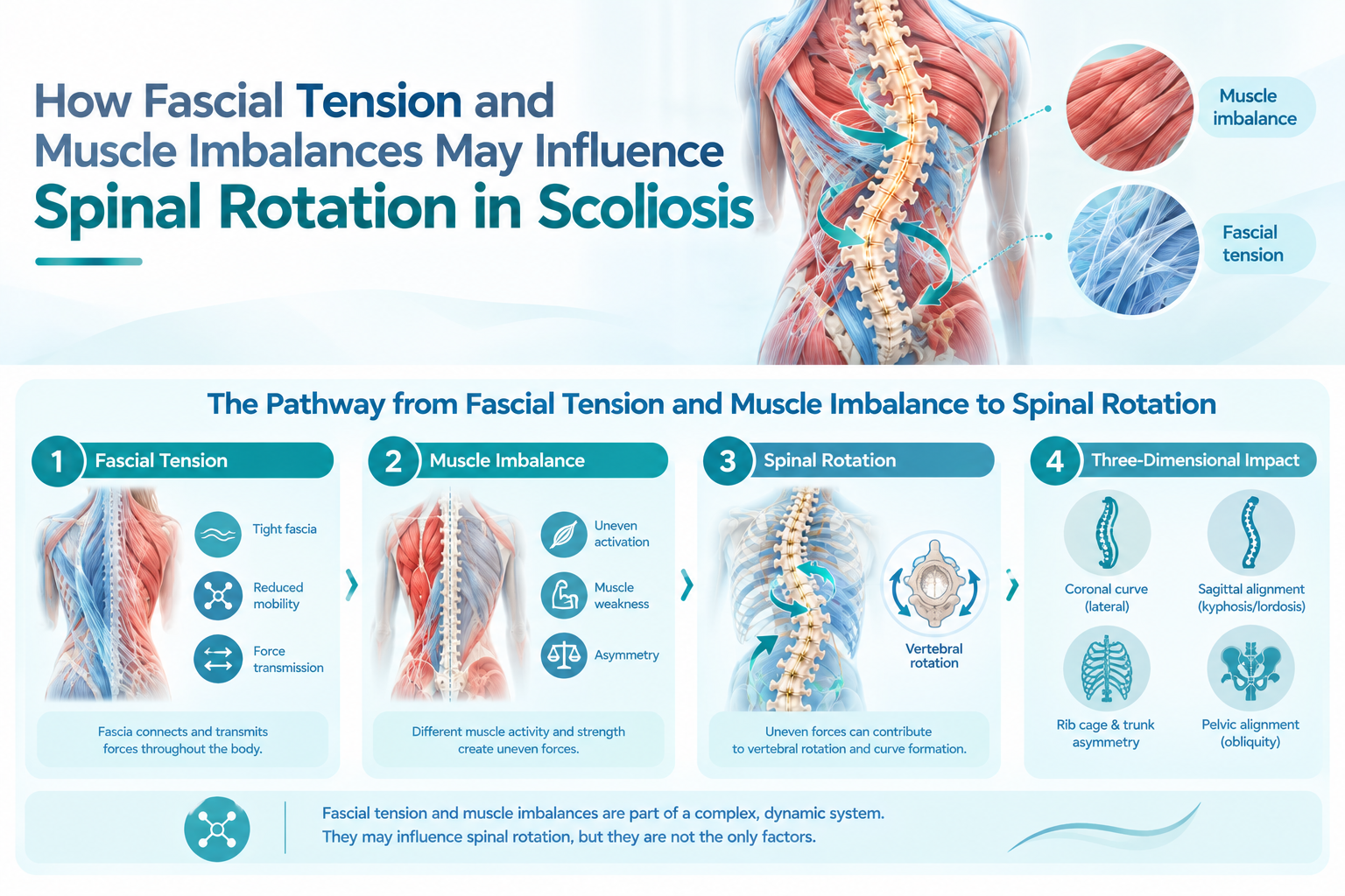
Learn how paraspinal muscle imbalance, fascial tension, and myofascial force transmission may influence spinal rotation in scoliosis, and why these factors should be considered within a three-dimensional biomechanical assessment.
Introduction
Scoliosis is more than a sideways curvature of the spine. It is a three-dimensional spinal deformity involving changes in the coronal plane, sagittal alignment, and axial rotation of the vertebrae. On a standing X-ray, scoliosis may appear primarily as a lateral curve, but the actual deformity can also involve vertebral rotation and changes in the shape and balance of the trunk. [1]
This three-dimensional nature helps explain why two people with similar Cobb angles can have noticeably different postures, rib prominence, shoulder asymmetry, pelvic alignment, or movement patterns.
One factor that has attracted increasing interest is the relationship between the spine and the surrounding muscles, fascia, and neuromuscular system. Paraspinal muscles do not simply support the spine. They continuously respond to spinal position, movement, loading, and changes in mechanical demand.
Research has identified differences in paraspinal muscle characteristics and activation between the convex and concave sides of scoliotic curves. However, an important question remains: Are these muscular differences a cause of spinal deformity, a consequence of it, or part of a continuing feedback loop? Current evidence does not support a simple answer. [2][3]
Understanding this relationship can nevertheless provide a useful biomechanical perspective on why spinal rotation may be accompanied by asymmetric muscle activity and altered soft-tissue tension.
---
What Does Spinal Rotation Mean in Scoliosis?
A normal spine is not perfectly flat in three dimensions. It has physiological curves in the sagittal plane and is capable of rotation around its longitudinal axis.
In scoliosis, however, abnormal rotation can occur together with the lateral curvature.
For example, a thoracic curve may be accompanied by:
- Rotation of the vertebral bodies
- Posterior prominence of the ribs on one side
- Asymmetric scapular position
- Altered thoracic shape
- Unequal paraspinal muscle activity
- Changes in trunk balance
This is why the Adams forward-bend test is useful during clinical assessment. When a patient bends forward, vertebral and rib-cage rotation can become more visible as a rib or lumbar prominence. [1]
Radiographic assessment remains essential because physical appearance alone cannot determine the complete structural characteristics of scoliosis. Standing spinal radiographs are generally used to evaluate the curve and calculate the Cobb angle, while additional imaging may be considered when clinically indicated. [1]
The Cobb angle is important, but it does not fully describe the three-dimensional nature of spinal deformity.
---
What Is Muscle Imbalance?
In the context of scoliosis, muscle imbalance generally refers to differences in muscle activity, strength, volume, morphology, endurance, or mechanical behavior between different regions or sides of the body.
The most frequently discussed muscles are the paraspinal muscles, including:
- Multifidus
- Erector spinae
- Longissimus
- Iliocostalis
- Other deep and superficial trunk stabilizers
These muscles contribute to spinal extension, rotation, segmental stabilization, and control of posture.
When the spine becomes asymmetrical, the mechanical demands placed on these muscles may also become asymmetrical.
A systematic review of paraspinal muscle characteristics in adolescent idiopathic scoliosis found low- to very-low-strength evidence for several differences between the convex and concave sides. Findings included differences in muscle fiber characteristics, muscle volume, muscle activity, atrophy, fatty infiltration, and gene expression. Importantly, the authors emphasized that these changes may be either a cause or a consequence of scoliosis. [2]
This distinction is critical.
It would be scientifically inaccurate to conclude that muscle imbalance alone causes idiopathic scoliosis.
---
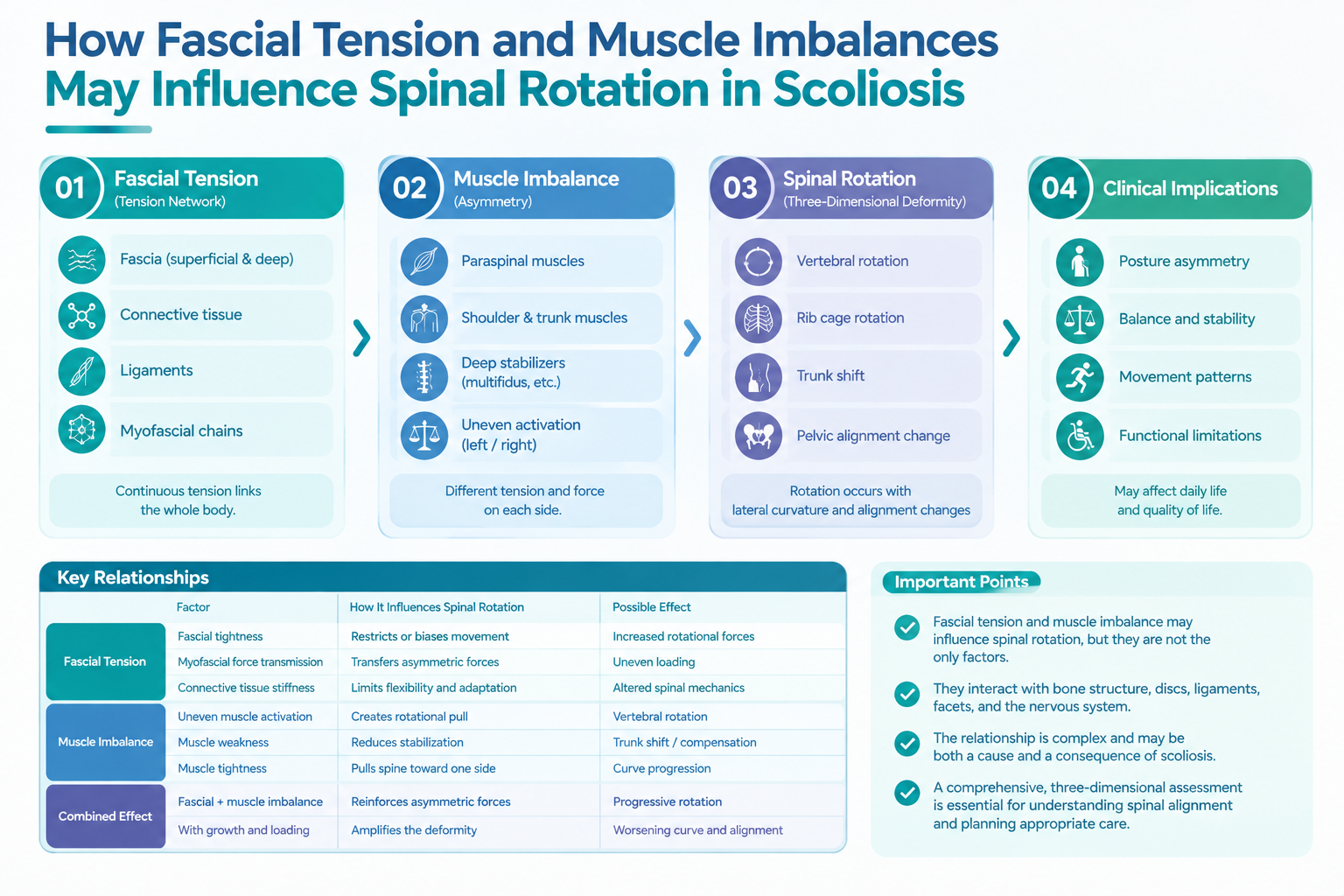
Paraspinal Muscle Asymmetry and Scoliosis
Electromyography (EMG) studies provide another way to examine the relationship between scoliosis and muscle activity.
A 2022 narrative review examined published surface EMG studies of adolescents with idiopathic scoliosis. Across the available measurements, some studies demonstrated greater activation on the convex side, particularly around the curve apex and during certain postural tasks, while many other measurements showed no significant side-to-side difference. [3]
This mixed evidence suggests that neuromuscular asymmetry is not necessarily uniform.
Muscle activation may vary according to:
- Curve location
- Curve magnitude
- Curve pattern
- Task being performed
- Body position
- Skeletal maturity
- Progression status
- Individual neuromuscular strategy
Therefore, a patient should not be assumed to have a simple pattern such as "tight muscles on one side and weak muscles on the other."
The neuromuscular system is considerably more complex.
---
How Muscle Activity May Influence Spinal Rotation
Muscles generate forces around the spine.
If these forces are symmetrical, they can contribute to balanced spinal stabilization. If forces become asymmetrical, however, they may contribute to differences in loading or movement across spinal segments.
Consider a simplified example.
If the paraspinal muscles on one side generate greater force than the corresponding muscles on the opposite side, the resulting mechanical moment may influence trunk position or rotation.
However, this should not be interpreted as evidence that asymmetric muscle activity independently creates structural scoliosis.
Instead, a more appropriate model is a dynamic interaction:
Spinal asymmetry → altered mechanical demand → altered muscle activation → altered loading → adaptation of movement and posture
This process may occur in the opposite direction as well:
Altered neuromuscular control → asymmetric loading → changes in spinal movement → potential contribution to asymmetrical posture
The relationship may therefore be bidirectional.
---
What Role Does Fascia Play?
Fascia is a connective-tissue system that surrounds, separates, and connects muscles and other anatomical structures.
Rather than viewing each muscle as an isolated unit, modern biomechanical research increasingly examines how mechanical forces can be transmitted between adjacent muscles and connective tissues.
This concept is often described as myofascial force transmission.
A systematic review of intermuscular force transmission found evidence that mechanical tension can be transferred between some anatomically connected muscular structures. However, the authors also emphasized that the functional significance of these connections remains incompletely understood and that differences in experimental methods make direct comparison difficult. [4]
Human in-vivo evidence has also been investigated.
A systematic scoping review identified studies supporting the possibility of myofascial force transmission in humans, but the researchers emphasized substantial heterogeneity and generally limited methodological quality. They therefore supported the existence of such force transmission only cautiously. [5]
This distinction is important for patients.
It is reasonable to say that fascia participates in mechanical force transmission.
It is not currently justified to say that:
"Tight fascia causes scoliosis."
The available evidence does not establish such a direct causal relationship.
---
Myofascial Chains and the Spine
Some biomechanical models describe the body as interconnected through longitudinal and functional myofascial pathways.
For example, anatomical and experimental research has investigated connections involving:
- The superficial backline
- Functional muscular chains
- Thoracolumbar fascia
- Paraspinal muscles
- Pelvic and lower-extremity musculature
These relationships provide a possible explanation for why movement or tension in one anatomical region can influence mechanical behavior elsewhere.
For example, the lumbar spine does not function independently of the pelvis and lower limbs. Likewise, thoracic spinal mechanics are influenced by the rib cage, shoulder girdle, abdominal wall, and breathing mechanics.
However, the existence of anatomical continuity does not automatically prove that a particular fascial chain causes scoliosis.
The distinction between anatomical connection, force transmission, and clinical causation must be maintained.
---
Fascial Tension and Rotational Forces
Spinal rotation is fundamentally a problem of three-dimensional mechanical balance.
When a vertebral segment rotates, surrounding tissues must accommodate that movement. These tissues include:
- Muscles
- Ligaments
- Fascia
- Intervertebral discs
- Facet joints
- Rib articulations
- Neural and connective tissues
If a scoliotic curve becomes structurally established, soft tissues may adapt to the altered geometry.
Some muscles may operate at shortened or lengthened positions. Others may change their activation strategy to maintain upright posture.
Over time, these adaptations may become part of the individual's habitual movement pattern.
This creates a potentially important feedback relationship:
Structural deformity → altered soft-tissue geometry → altered muscle activation → altered movement → further adaptation
The precise contribution of each component probably varies considerably between individuals.
---
Structural Rotation vs. Functional Rotation
One of the most important distinctions is between structural spinal rotation and rotation associated primarily with functional or postural asymmetry.
A functional asymmetry may change with:
- Position
- Movement
- Muscle activation
- Leg or pelvic position
- External loading
A structural deformity is more persistent and is evaluated using clinical examination and imaging.
This distinction matters because an intervention that improves posture or muscle coordination does not necessarily mean that an established vertebral deformity has been anatomically corrected.
For this reason, a patient's subjective improvement in posture, movement, or discomfort should not automatically be equated with a reduction in Cobb angle or structural correction.
---
The Tensegrity Perspective
The relationship between muscles, fascia, and spinal alignment can also be considered through the broader concept of biotensegrity.
In a tensegrity-inspired model, stability is produced not simply by rigid structures stacked on top of one another but through a balance between tension and compression.
Applied conceptually to the spine, this model encourages clinicians and researchers to consider:
- Distributed mechanical forces
- Muscle tension
- Connective-tissue continuity
- Segmental stability
- Global posture
- Dynamic adaptation
This can be useful as a biomechanical framework.
However, tensegrity should be regarded as a conceptual model, not as an established diagnosis or proven explanation for idiopathic scoliosis.
Modern spinal deformity research increasingly uses sophisticated radiographic measurements, three-dimensional imaging, biomechanical modelling, and alignment analysis to understand spinal deformity more comprehensively. [6]
---
Can Fascial or Muscle-Based Treatment Correct Scoliosis?
This question requires particular caution.
Exercise and rehabilitation can play an important role in scoliosis management, particularly when they are individualized and integrated into an appropriate clinical treatment plan.
Scoliosis-specific exercise approaches such as Schroth-based programs have been investigated in systematic reviews and meta-analyses, with evidence suggesting potential improvements in outcomes such as Cobb angle, trunk rotation, and quality of life in selected patients. However, study quality and protocols vary, and exercise should not be presented as universally capable of reversing structural scoliosis. [7]
Similarly, research on fascial manipulation is growing, but a 2024 scoping review concluded that the evidence base remains limited and that further randomized controlled trials are needed. [8]
Therefore, statements such as:
- "Fascial release cures scoliosis."
- "Muscle imbalance is the primary cause of idiopathic scoliosis."
- "Releasing one fascial chain will straighten the spine."
go beyond what current evidence can establish.
A more scientifically appropriate statement is:
Muscle and fascial interventions may influence movement, pain, posture, and neuromuscular control, but their ability to modify established structural scoliosis requires further evidence.
---
A Pattern-Based Approach to Scoliosis
Because scoliosis is highly variable, some clinicians have explored classification systems that go beyond the Cobb angle.
A pattern-based approach may consider:
- Curve direction
- Curve location
- Vertebral rotation
- Pelvic alignment
- Shoulder and trunk asymmetry
- Muscle activation
- Functional movement patterns
- Postural compensation
One clinician-developed approach is presented in Scoliosis, Meridian-Based Isometric Exercises and JESU5C Pattern Suit, which describes the Hyun-Ga Manual Technique (HGMT), meridian-based isometric exercises, and the JESU5C pattern classification for idiopathic scoliosis. [9]
Such a framework should be understood as a clinical approach or hypothesis-generating model, rather than as an established replacement for conventional radiographic classification or evidence-based scoliosis assessment.
---
Why the Muscle–Fascia–Spine Relationship Matters
Even when muscle imbalance or fascial tension is not the primary cause of scoliosis, understanding these tissues can still be clinically useful.
The spine is a living, moving system.
A patient with scoliosis may develop adaptations in:
- Muscle recruitment
- Balance
- Gait
- Breathing mechanics
- Shoulder movement
- Pelvic control
- Trunk stabilization
These adaptations may influence symptoms and functional capacity even when the structural curve itself remains relatively stable.
This is why scoliosis management increasingly considers more than simply the number of degrees measured on an X-ray.
The Cobb angle remains essential, but a comprehensive assessment may also consider:
Cobb angle + vertebral rotation + sagittal alignment + coronal balance + posture + neurological findings + skeletal maturity + symptoms + functional movement
Together, these factors provide a more complete picture.
---
What Patients Should Know
If you have scoliosis, it is important not to assume that muscle tightness or fascial tension is the cause of your condition.
Idiopathic scoliosis means that a specific underlying cause has not been identified. The Scoliosis Research Society notes that most scoliosis cases are idiopathic and that multiple biological and mechanical factors may be involved. [1]
Patients should also be cautious about treatment claims that promise rapid structural correction without appropriate clinical and radiographic assessment.
A qualified clinician should consider:
- The Cobb angle
- Curve location and pattern
- Vertebral rotation
- Sagittal alignment
- Coronal balance
- Skeletal maturity
- Neurological findings
- Pain and functional limitations
- Evidence of curve progression
- The patient's overall treatment goals
If significant neurological symptoms, severe pain, rapid progression, or other unusual findings are present, appropriate medical evaluation is particularly important.
---
Conclusion
Muscle imbalance and fascial tension provide an interesting perspective on the biomechanics of scoliosis.
Research demonstrates that paraspinal muscle characteristics and neuromuscular activation can differ between the sides of a scoliotic curve, while biomechanical studies suggest that forces can be transmitted through interconnected muscular and fascial structures. [2][3][4][5]
However, these findings do not prove that muscle imbalance or fascial tension is the primary cause of idiopathic scoliosis.
A more useful interpretation is that the spine, muscles, fascia, and nervous system may participate in a continuing process of mechanical adaptation.
For patients and clinicians, this perspective encourages a broader understanding of scoliosis: not simply as a lateral curve measured by the Cobb angle, but as a three-dimensional and dynamic spinal condition involving structure, movement, neuromuscular control, and whole-body alignment.
Future research combining three-dimensional imaging, electromyography, musculoskeletal modelling, and longitudinal clinical data may help clarify how these systems interact—and which interventions can meaningfully influence spinal function and structural progression.
---
References
[1] Scoliosis Research Society. Scoliosis. Scoliosis Research Society.
[2] Associations between paraspinal muscle characteristics and spinal curvature in conservatively treated adolescent idiopathic scoliosis: a systematic review. European Spine Journal. 2023.
[3] Ng, P. T. T., Claus, A., Izatt, M. T., Pivonka, P., & Tucker, K. Is spinal neuromuscular function asymmetrical in adolescents with idiopathic scoliosis compared to those without scoliosis?: A narrative review of surface EMG studies. Journal of Electromyography and Kinesiology. 2022.
[4] Krause, F., Wilke, J., Vogt, L., & Banzer, W. Intermuscular force transmission along myofascial chains: a systematic review. Journal of Anatomy. 2016.
[5] Ajimsha, M. S., Shenoy, P. D., Surendran, P. J., Jacob, P., & Bilal, M. J. Evidence of in-vivo myofascial force transfer in humans—A systematic scoping review. Journal of Bodywork & Movement Therapies. 2022.
[6] Smith, J. S., Shaffrey, C. I., Bess, S., et al. Recent and Emerging Advances in Spinal Deformity. Neurosurgery. 2017.
[7] Ceballos-Laita, L., Carrasco-Uribarren, A., Cabanillas-Barea, S., et al. The effectiveness of Schroth method in Cobb angle, quality of life and trunk rotation angle in adolescent idiopathic scoliosis: a systematic review and meta-analysis. European Journal of Physical and Rehabilitation Medicine. 2023.
[8] Isaji, Y., Sasaki, D., Kon, Y., Kurasawa, Y., & Kitagawa, T. Fascial manipulation for musculoskeletal disorders: A scoping review. Journal of Bodywork & Movement Therapies. 2024.
[9] LEE, Yongsub, & Lee, Minsu (translator). Scoliosis, Meridian-Based Isometric Exercises and JESU5C Pattern Suit: “JESU5C” a New Classification of Idiopathic Scoliosis and HGMT (Hyun-Ga Manual Technique). Amazon Kindle Edition.